Versions Compared

Key

  • This line was added.
  • This line was removed.
  • Formatting was changed.

...

Their Laboratories

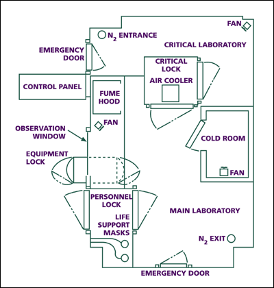
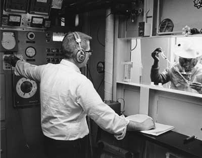
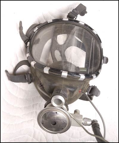
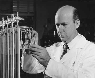
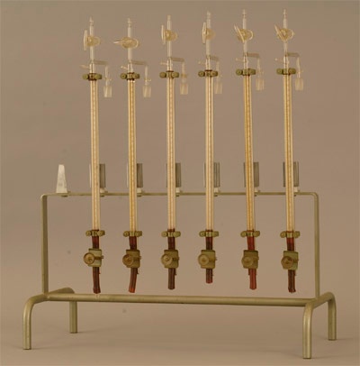
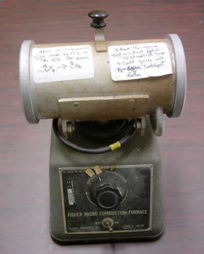
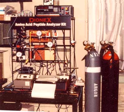
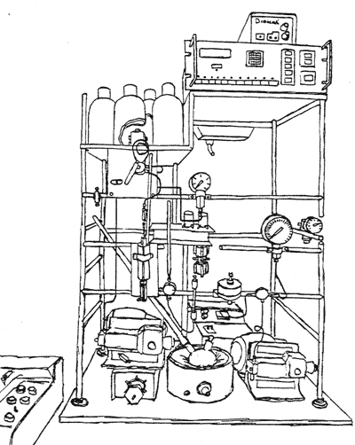
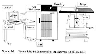
At NIH, Thressa and Earl have worked in the same building, sharing common facilities and instruments. Yet each has maintained her or his laboratory space separately as an independent researcher. A good example of a facility used in common is the anaerobic laboratory in which they have conducted various experiments in oxygen-free conditions. Standard pieces of equipment for biochemical research, such as the centrifuge and the Warburg apparatus, are found in each laboratory. Here is a selected list of facilities and instruments in their laboratories over time.

Table of Contents
maxLevel2
minLevel2

Div
classusagrid-row grid-gap
Div
classusadesktop:grid-widthcol-two-thirds8

Anaerobic Laboratory-An NIH First

In 1967, the first anaerobic laboratory for biomedical research was built in Building 3 on the NIH's Bethesda campus. This quarter of a million dollar facility demonstrated the NIH's strong commitment to the research programs led by Thressa and Earl Stadtman. Thressa's research on methane biosynthesis and selenium biochemistry particularly benefited from this unique facility. Until the closing of Building 3 in 2002, the anaerobic laboratory served numerous researchers at NIH as well as visiting scientists from around the world.The anaerobic laboratory was furnished with the usual laboratory equipment, but it was filled with a mixture of nitrogen and hydrogen, instead of ordinary air. An "oxygen-free" atmosphere is crucial in dealing with bacteria that are killed in the presence of oxygen or with biological compounds that are inactivated when exposed to air. This unique facility allowed researchers to conduct multi-step experiments with various instruments, including manipulations that were extremely difficult in conventional anaerobic "glove boxes." Because of the danger of working in an anaerobic atmosphere, a researcher must wear a special mask fitted with an air-delivery tube while an observer outside monitors a two-way communication system.

Video: Michael Poston on the anaerobic laboratory.

Running time: 3:00 minutes 
Small (56k modem)
Large (ISDN/DSL)

Div
classusadesktop:grid-widthcol-one-third4

Floor plan of the anaerobic laboratory.
Floor plan of the anaerobic laboratory

Inside the anaerobic laboratory.
Inside the anaerobic laboratory

Respirator mask used iside the anaerobic laboratory Fermenter Room

...

Running time: 2:10 minutes
Small (56k modem)
Large (ISDN/DSL)

Div
classusagrid-row grid-gap
Div
classusadesktop:grid-widthcol-two-thirds8

Warburg Apparatus

The Warburg apparatus is an analytical instrument for measuring the pressure of gases and vapors from biochemical reactions. It was named after its inventor, the German biochemist Otto Heinrich Warburg (1883-1970). Warburg pioneered research on cell respiration and tumor metabolism, work for which he won the Nobel Prize in Physiology or Medicine in 1931. Originally, the Warburg apparatus was used to study "respiration" or the uptake of gaseous oxygen and the production of carbon dioxide by various cells or tissues. Later, it also proved useful in examining "fermentation," a process of breaking down organic compounds in the absence of oxygen. It was an ideal instrument for both aerobic and anaerobic enzyme studies until it was replaced by various spectrometers.

The Warburg apparatus is based on the principle that, at constant temperature and constant gas volume, any changes in the amount of a gas can be measured by changes in its pressure. As a closed system, it consists of a detachable flask equipped with one or more sidearms for additions of chemicals and a manometer (pressure gauge) containing a liquid of known density. The sample of interest is placed in the main chamber and the flask is immersed in a constant temperature water bath. It is then shaken to facilitate rapid gas exchange between the fluid and the gas phase. At equilibrium a starting reference point is read on the manometer, and then the reaction is started. The volume of gas produced or absorbed is determined at specific time intervals.

Earl used this apparatus for his early research on fatty acid metabolism and also for his research on aging in the 1980s. It was also an important tool for Thressa in studying the role of vitamin B12 -dependent enzymes in methane biosynthesis and selenium biochemistry.

Div
classusadesktop:grid-widthcol-one-third4

Earl using the Warburg apparatus, 1952.

The Warburg apparatus.

Div
classusagrid-row grid-gap
Div
classusadesktop:grid-widthcol-two-thirds8

Micro-Combustion Furnace

This micro-combustion furnace, manufactured around 1942 by the Fisher Scientific and Eimer & Amend company, was used for Thressa's research on methane fermentation in the 1950s and 1960s. Radioactive methane was oxidized to radioactive carbon dioxide by a catalyst in a tube heated in the furnace. In this way, a given amount of radioactive methane could be determined quantitatively. Later on other direct methods of measuring methane using special chromatographic equipment became available.

The furnace uses an electrical current to heat substances placed in a tube. The heating coils are controlled by a variable transformer, which converts the electrical current into heat inside the furnace so that no heat is lost. The electrical current input-and therefore the temperature-is controlled by the dial on the base of the instrument. The furnace can produce temperatures ranging from 200o -1,700oF.

Div
classusadesktop:grid-widthcol-one-third4

Fisher Scientific and Eimer and Amend Micro-Combustion Furnace.

Div
classusagrid-row grid-gap
Div
classusadesktop:grid-widthcol-two-thirds8

Amino Acid/Peptide Analyzer

The amino acid/peptide analyzer was sold as a kit by the Dionex Corporation. In 1980, Thressa's research assistant, Joe Davis, and her postdoctoral fellow, Greg Dilworth, assembled it, like an erector set, by attaching equipment to a frame of connected rods on a work cart in the laboratory. They used the analyzer for identifying amino acids in selenium-containing proteins. Earl's group also took advantage of it for analyzing amino acids in oxidized proteins. These days, high pressure liquid chromatography (HPLC) systems are available from many manufacturers, and any of them can be configured to do the job of analyzing amino acids and peptides.

The instrument consists of several parts. First, it is controlled by a Dionex Chromatograph Programmer, a controlling computer, which programs the times at which different solvents are pumped and the temperatures at which they are pumped. Second, solvents are kept in glass bottles on top of the rack, and their temperature is usually kept at 40o to 65oF by the Dionex Column Heater Control. Third, connected to the mixing column is the filtering and detecting device, the Gilson Spectra/flo Fluorometer. Finally, all the information-the pH level of solvents and the program of solvents/temperatures/time is recorded in the Shimadzu Chromatopac C-R3A Recorder. This particular recorder was a later addition to the instrument.

The operation of the amino acid/peptide analyzer is largely automated but requires some experience. A mixture of amino acids is injected into the system and pumped into the mixing column. Next, each amino acid is then eluted or washed out from the column in a specific order. This is accomplished by pumping in solvents of increasing pH and by varying the column temperature. The eluted amino acid in buffer is then mixed with the detecting materials at the bottom of the column. Subsequently, the mixture goes into the detecting device, in which the filter controls the specificity of the fluorometer. As it passes through the filter, the emission of fluorescence is detected.

Div
classusadesktop:grid-widthcol-one-third4


The instrument set up in Thressa's laboratory.


Drawing of the amino acid/peptide analyzer.

Div
classusagrid-row grid-gap
Div
classusadesktop:grid-widthcol-two-thirds8

Electron Paramagnetic Resonance (EPR)

Electron Paramagnetic Resonance (EPR) is an instrument that detects substances with a particular magnetic property called "paramagnetism." Paramagnetism can be found in such metal atoms as aluminum and platinum, or such metal ions as Fe3+ and Cu2+, which become magnetized in a magnetic field but lose their magnetic power when the field is removed. This property is partly due to the "spin" movement of electrons. The electron, one of the fundamental particles that constitute an atom, orbits around the atom's nucleus. But it also spins by itself, like a spinning top. Only two directions of spin, opposite to each other, are allowed. When two electrons of different spin properties are paired, the effect of spinning-mathematically represented by the angular momentum-is cancelled out. However, when there is an unpaired electron in the substance, the spin effect can be detected. Hence EPR is also called Electron Spin Resonance (ESR). Since it is one of the most sensitive instruments that can identify substances having unpaired electrons, EPR has been successfully applied in various fields of study, such as detecting impurities in semi-conductors. In biochemistry, it has become a powerful tool for finding "free radicals" in cells. For this reason, Earl and his co-workers began to use it in 1988. After updating the first model installed at that time (the Bruker ESP 300) several times, the laboratory has purchased two more advanced models, Elexsys E580 and EMX, which are now installed in Building 50. Recently, Thressa's group also has taken advantage of this instrument in research on enzymes that contain selenium as well as molybdenum (a heavy metal).

The most distinctive feature of EPR is the two big magnets. The sample is placed in the microwave resonator located in the gap between the magnets. When a strong magnetic field is applied to the sample, a small magnetic moment arising from the spin of an unpaired electron is oriented in a direction either parallel or anti-parallel to the applied field. This creates a unique energy level for the sample in the given magnetic field. Then, EPR's microwave generator sends a wave of a specific energy (measured in frequency) to the microwave resonator where the sample is placed. When the microwave's energy (i.e., its frequency) matches exactly with the energy level, it is absorbed by the sample. This "energy-absorption" or "frequency-matching" phenomenon is called "resonance." For technical reasons, EPR detects resonance at a fixed microwave frequency by changing or scanning the strength of the magnetic field. With the positions of resonance lines and their amplitudes, it is possible to determine what kind of free radicals are in the sample and how much of them there is. In addition, since EPR's resonance lines are split further according to the overall environment for electrons-in particular, the presence of nuclei nearby-the analysis of this splitting can also provide useful information on the structural identity of the sample.

Video: Moon Bin Yim on electron paramagnetic resonance.
Running time: 1:17 minutes 
Small (56k modem)
Large (ISDN/DSL)

Div
classusadesktop:grid-widthcol-one-third4

Drawing of EPR.
Drawing of EPR.

Div
classusagrid-row grid-gap
Div
classusadesktop:grid-widthcol-two-thirds8

Spectrophotometer and High Pressure Liquid Chromatography (HPLC)

Since the early 1980s, Earl and his co-workers have used various analytical methods to measure the level of oxidized, and thus inactivated, enzymes for their research on protein degradation and aging. For example, the spectrophotometer, which measures the relative intensities of light in different parts of the spectrum, can detect changes in the level of oxidized proteins.

In separating and identifying oxidized segments of proteins (i.e., oxidized peptides or amino acids), various chromatographic techniques are useful. Among them is the high pressure liquid chromatography commonly known as HPLC. Its operational principle is simple: the sample of interest is injected into HPLC's column, and as the mobile phase passes through the column, the different products are separated and detected. In Earl's laboratory, a variety of columns are used to separate and identify compounds, depending on their molecular properties, such as size, ionic charge, and affinity to specific molecules. In Thressa's research, HPLC separation of selenium containing proteins, peptides and amino acids is important.

Div
classusadesktop:grid-widthcol-one-third4

Spectrophotometer
Spectrophotometer

High Pressure Liquid Chromatograph.