As the field researchers gathered many species of ticks and ticks in many stages of their life cycle, Dr. Robert Cooley developed a huge collection of them. Cooley was the head entomologist at Canyon Creek Schoolhouse laboratory. This collection would help later researchers solve questions about other tick-borne diseases. In this photo, Cooley shows off his world-class tick collection kept in jars in a huge card file cabinet. The photo was taken in Building One (which opened in 1928), not the Canyon Creek Schoolhouse laboratory, on December 3, 1946. Cooley had been developing this collection since his first days studying RMSF forty years before.
Div
class
desktop:grid-col-6
Image Modified
Span
id
credit
class
credit
Image: Wikimedia Commons
Div
class
grid-row grid-gap
Div
class
desktop:grid-col-6
How do you get RMSF?
Occurrence
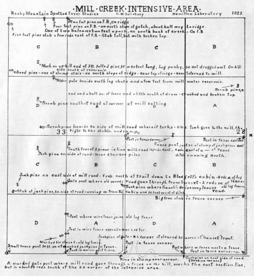
One way of finding out how a disease is spread is by looking at where it occurs. Rocky Mountain spotted fever came to be studied in Montana’s Bitterroot Valley because the bites of the western slope’s ticks caused a particularly deadly infection, meaning that more people died. By mapping and charting where they found ticks or where people got RMSF, the researchers could find the ticks and animals they needed to study. Mapping turned up interesting insights, such as one area may have a heavily infected tick population, but an area across a stream did not. Ticks do not like to swim.
Div
class
desktop:grid-col-6
Image Modified
C.M. Salisbury drew this grid map of an area of land near Mill Creek in 1922. Included were the sites of stumps, trees, and roads. The researchers could use it to keep track of where they collected ticks.
Span
id
credit
class
credit
Image: Rocky Mountain Laboratories, 961b
...
Div
class
grid-row grid-gap
Div
class
desktop:grid-col-6
6. Feed the adult ticks on an infected guinea pig. Then eviscerate the ticks and grind them in a mortar for 10-15 minutes with sterile sand and a bit of salt solution. This will separate their internal organs from their exoskeletons to create an emulsion.
7. Dilute the emulsion with salt solution so 1 cubic centimeter of emulsion equals two or more tick viscera.
8. Test the emulsion on two guinea pigs to find the minimal infectious dose. Both guinea pigs have to get RMSF to consider the emulsion for the vaccine.
9. Dilute the emulsion again to equal one tick. Add phenol so that the final product has 0.5% phenol (preservative-disinfectant).
Div
class
desktop:grid-col-6
Image Modified
Span
id
credit
class
credit
Image: Rocky Mountain Laboratories, 746
...
Div
class
grid-row grid-gap
Div
class
desktop:grid-col-6
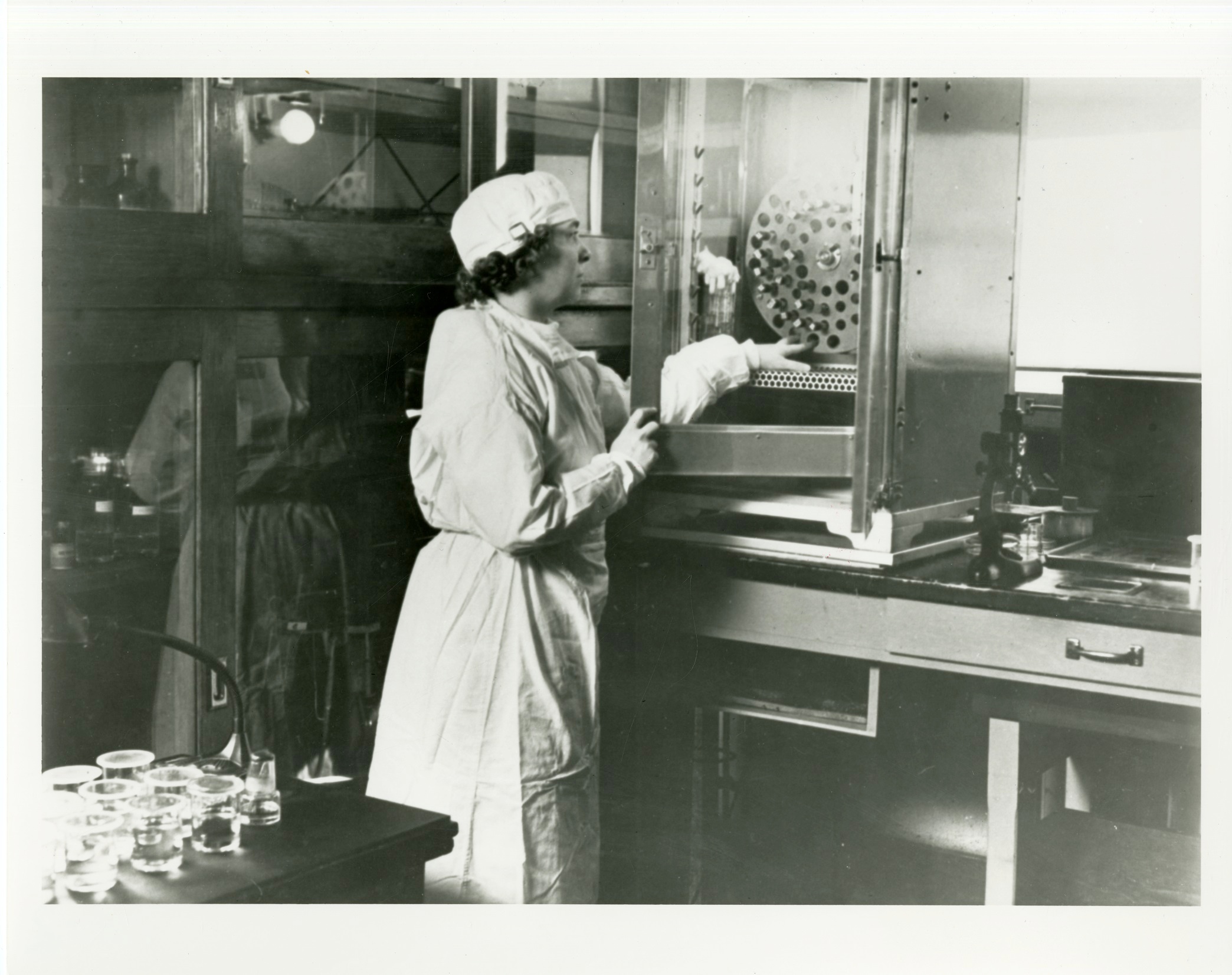
11. Use a centrifuge to separate the precipitate, as Emily Emmart is doing in the photo, because it doesn’t go through filter paper, and the vaccine’s potency is destroyed by a Berkefeld filter. The supernatant fluid (clear fluid) is the vaccine.
Div
class
desktop:grid-col-6
Image Modified
Span
id
credit
class
credit
Image: National Library of Medicine, 101447516
...
Div
class
grid-row grid-gap
Div
class
desktop:grid-col-6
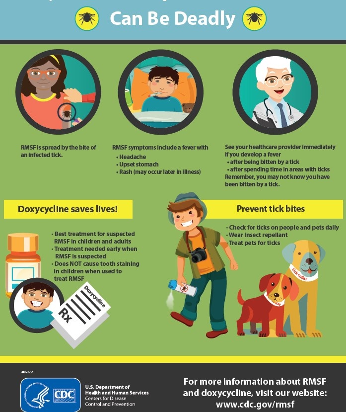
A Vaccine Made Unnecessary
One question not answered at the Canyon Creek Schoolhouse laboratory was if there was an effective treatment for Rocky Mountain spotted fever. But in the late 1940s, antibiotics were found to cure the disease; this discovery made the Spencer-Parker vaccine obsolete. This current infographic from the CDC reminds us that RMSF is still a threat to people’s health. Visit their website for more information https://www.cdc.gov/rmsf/index.html