
...
| Div | ||||||||||
|---|---|---|---|---|---|---|---|---|---|---|
| ||||||||||
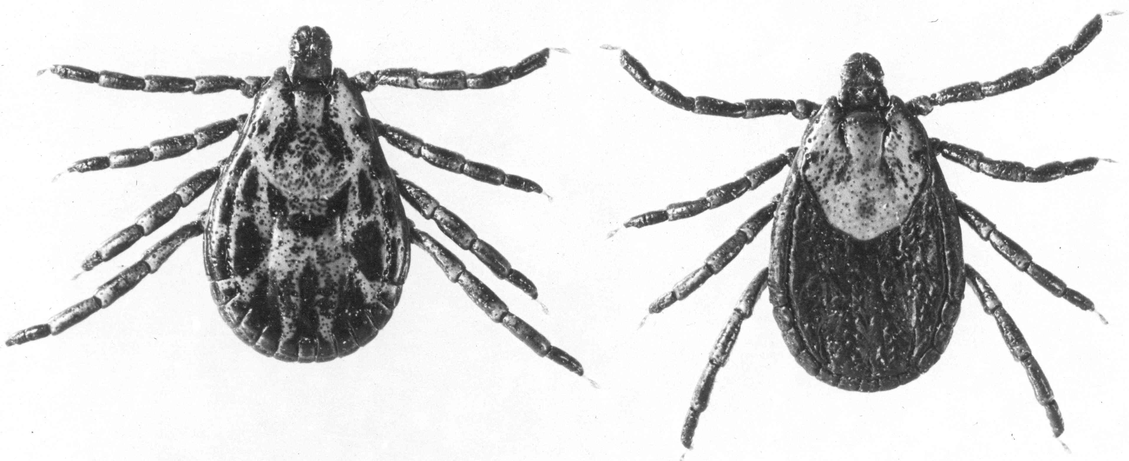
In spring, the nymphs once again attach to an animal and feed, drop to the ground, and digest their meals. This time they molt to adults, like these Rocky Mountain wood ticks, Dermacentor andersoni. They spend their second winter as unfed adults.
The next spring they climb grass and bushes to attach to animals.
But as adults, ticks mate as well as feed. This is when the ticks can get the Rickettsia rickettsii bacteria from the animals they feed on. Even though the bacteria don’t hurt the ticks, it infects all of their cells, and is passed on through the egg. The next generation of ticks will be infected. Images: Office of NIH History and Stetten Museum, 14541554-58 |


| Div | ||||||||||
|---|---|---|---|---|---|---|---|---|---|---|
| ||||||||||
|


| Div | ||||||||||
|---|---|---|---|---|---|---|---|---|---|---|
| ||||||||||
|


| Div | ||||||||||
|---|---|---|---|---|---|---|---|---|---|---|
| ||||||||||
How do you know you have RMSF?
|

| Div | |||||||||||
|---|---|---|---|---|---|---|---|---|---|---|---|
| |||||||||||
Can we prevent RMSF?
|


| Div | ||||||||||
|---|---|---|---|---|---|---|---|---|---|---|
| ||||||||||
Vaccine
|


| Div | ||||||||||
|---|---|---|---|---|---|---|---|---|---|---|
| ||||||||||
|

| Div | ||||||||||
|---|---|---|---|---|---|---|---|---|---|---|
| ||||||||||
|


| Div | ||||||||||
|---|---|---|---|---|---|---|---|---|---|---|
| ||||||||||
|


| Div | ||||||||||
|---|---|---|---|---|---|---|---|---|---|---|
| ||||||||||
|


...
| Div | ||||||||||
|---|---|---|---|---|---|---|---|---|---|---|
| ||||||||||
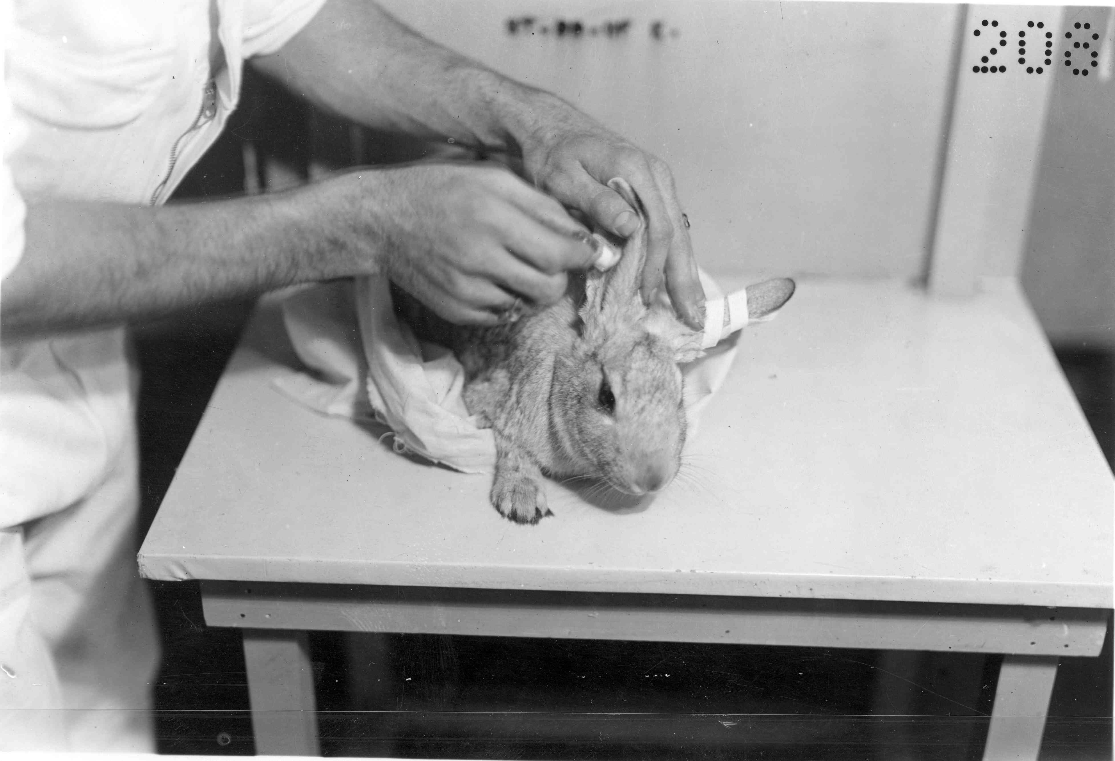
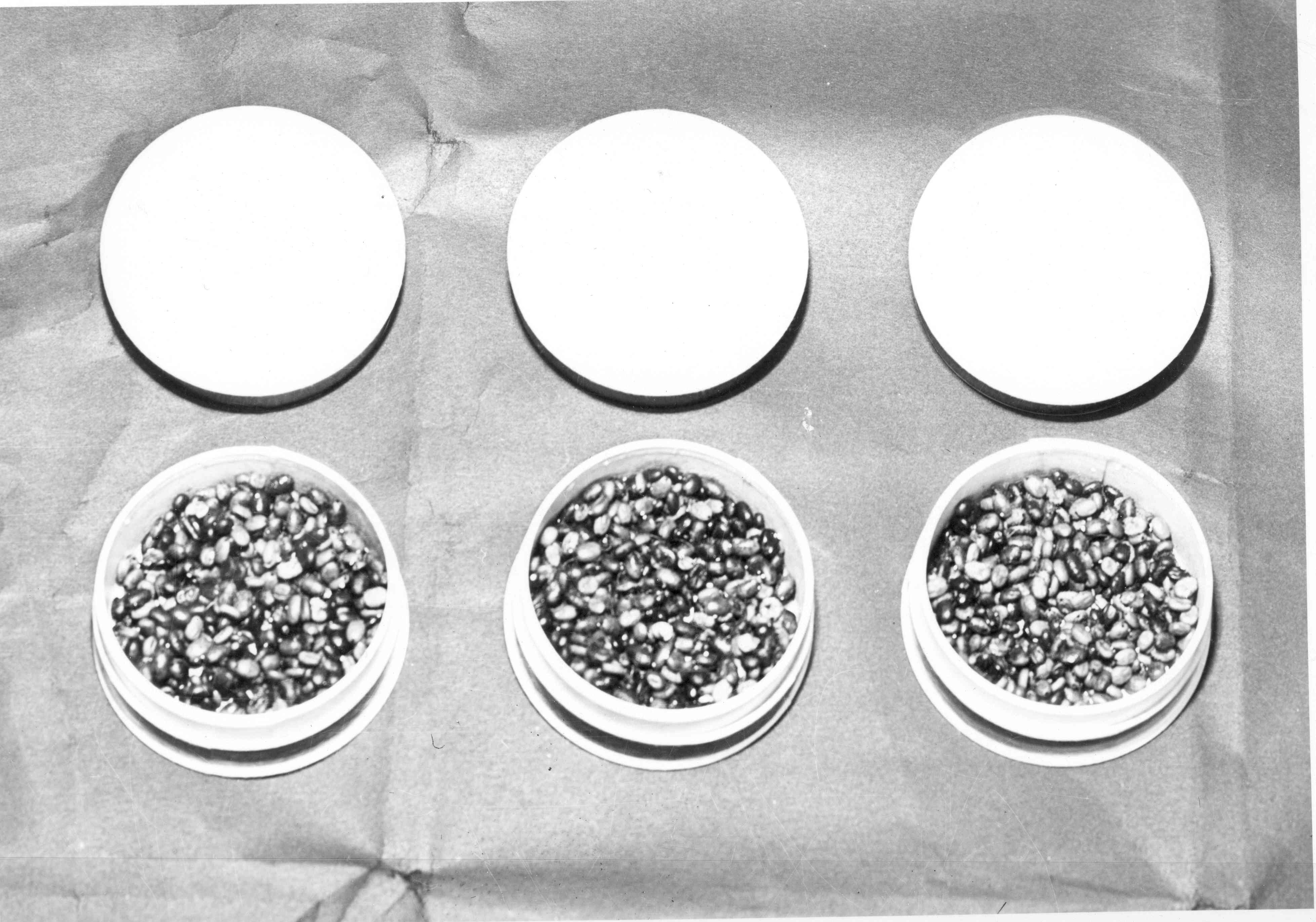
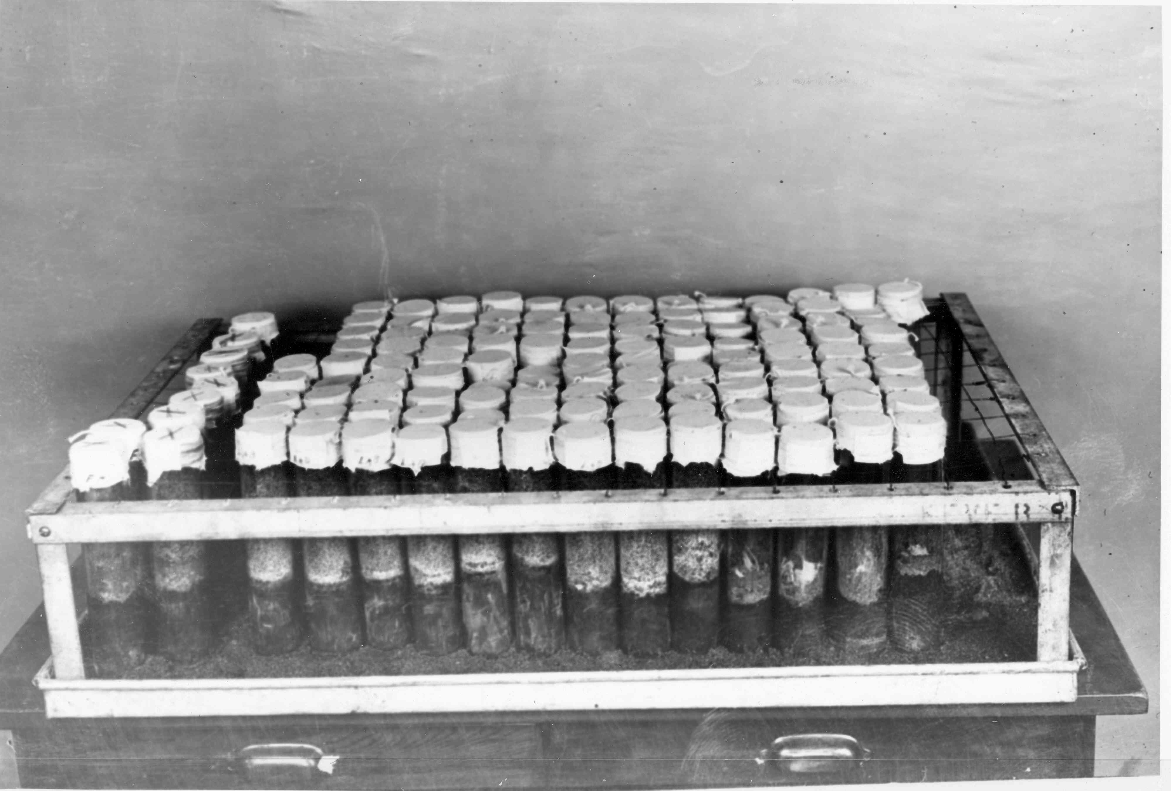
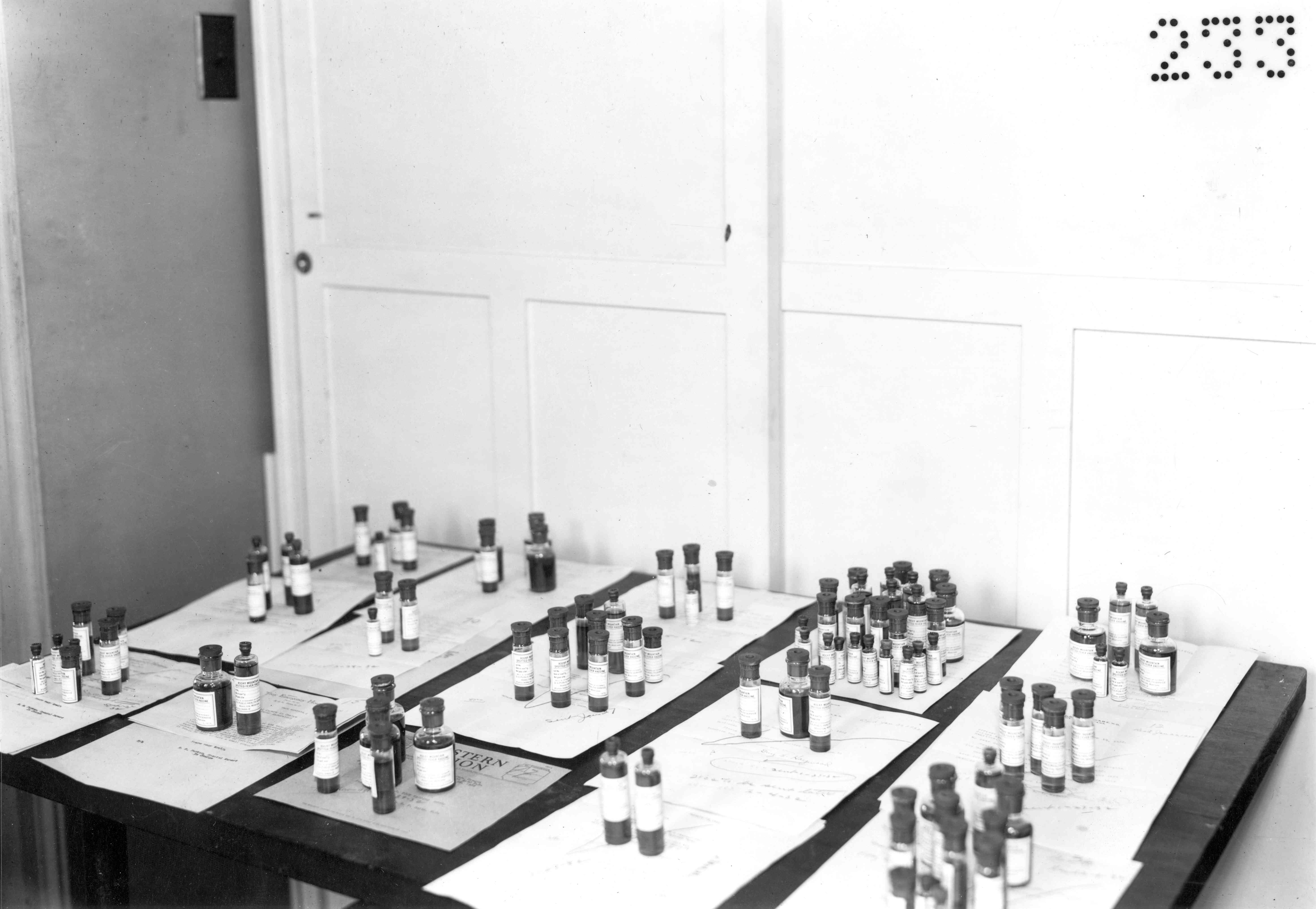
Vaccine Production StepsIn 1925, Drs. Roscoe Spencer and Ralph Parker described their method for creating RMSF vaccine. The method would become more streamlined and automated after they moved into the Building One laboratory in 1928 and got better space and equipment than the Canyon Creek Schoolhouse laboratory could provide. These photos include some taken at Building One after the laboratory moved from the schoolhouse.
|

| Div | ||||||||||
|---|---|---|---|---|---|---|---|---|---|---|
| ||||||||||
|

| Div | ||||||||||
|---|---|---|---|---|---|---|---|---|---|---|
| ||||||||||
|

...
| Div | ||||||||||
|---|---|---|---|---|---|---|---|---|---|---|
| ||||||||||
|

...
| Div | ||||||||||
|---|---|---|---|---|---|---|---|---|---|---|
| ||||||||||
|

...
| Div | ||||||||||
|---|---|---|---|---|---|---|---|---|---|---|
| ||||||||||
|

| Div | ||||||||||
|---|---|---|---|---|---|---|---|---|---|---|
| ||||||||||
|

...
Overview
Content Tools
ThemeBuilder