
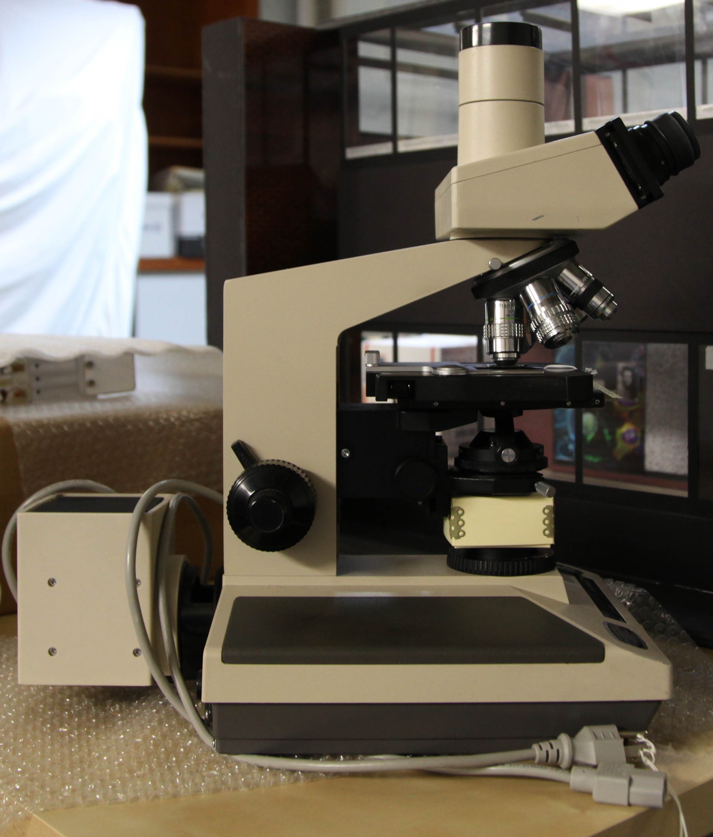
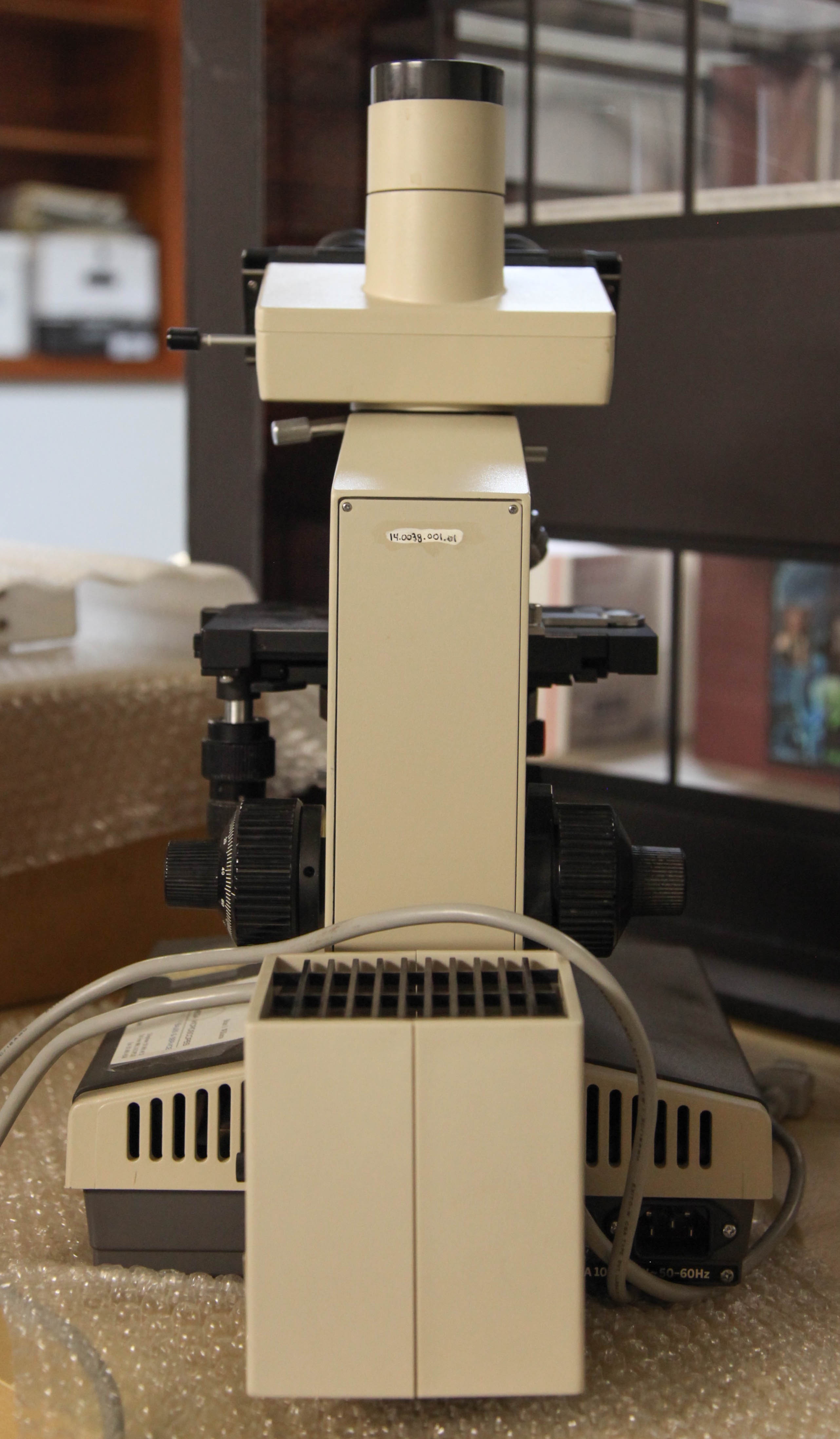
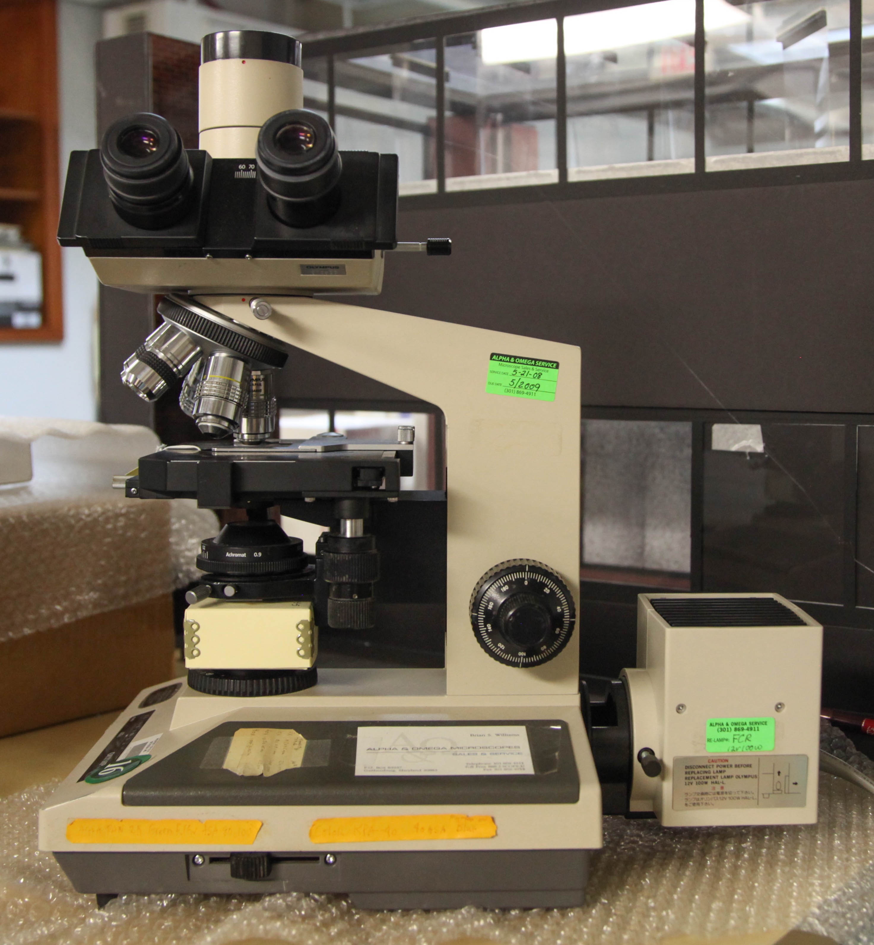
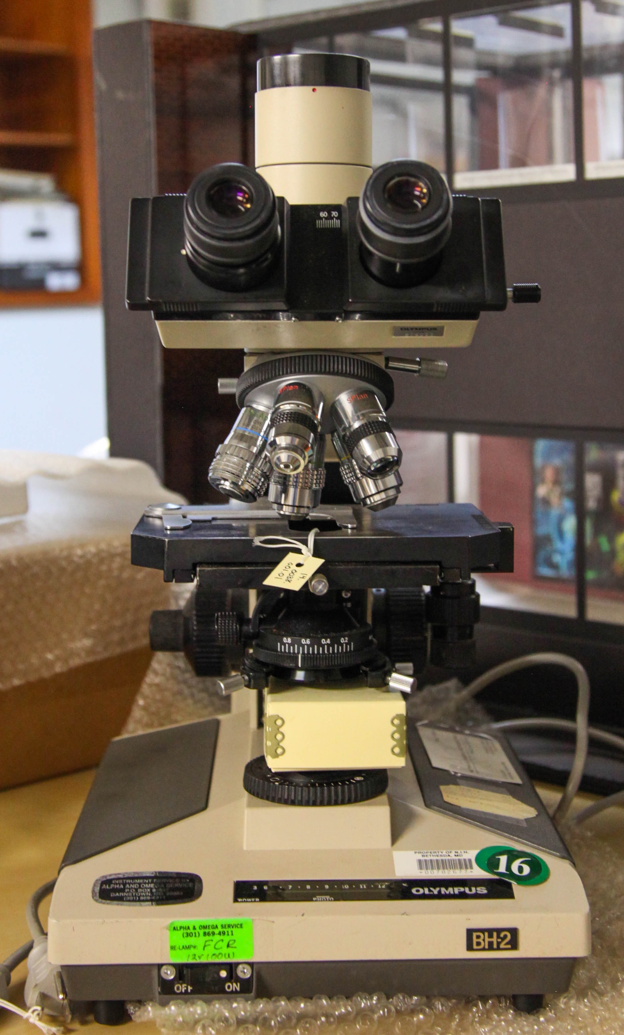
Olympus BH-2 Microscope, c. 1985
14.0038.001
“He would jump up from the scope, pulling his hair, and get all excited about a number of different experiments.”
- —Dr. Gary Jones, NCI
| Dive | |||||||||||||||||||
|---|---|---|---|---|---|---|---|---|---|---|---|---|---|---|---|---|---|---|---|
| |||||||||||||||||||
|
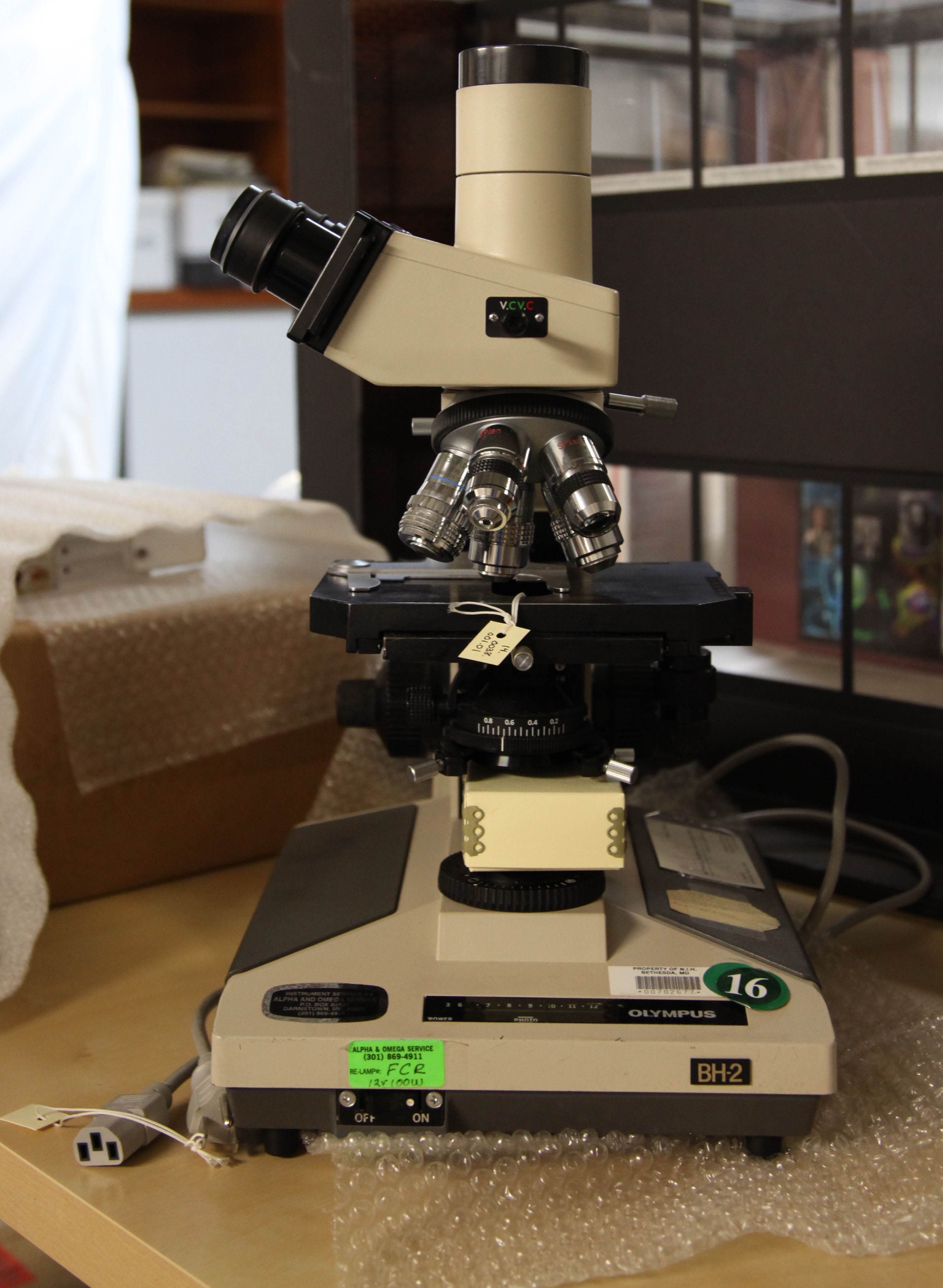
Learn more about the BH-2 in this Olympus Microscope manual in pdf format, hosted on Alan Wood's website (7.43 MB)
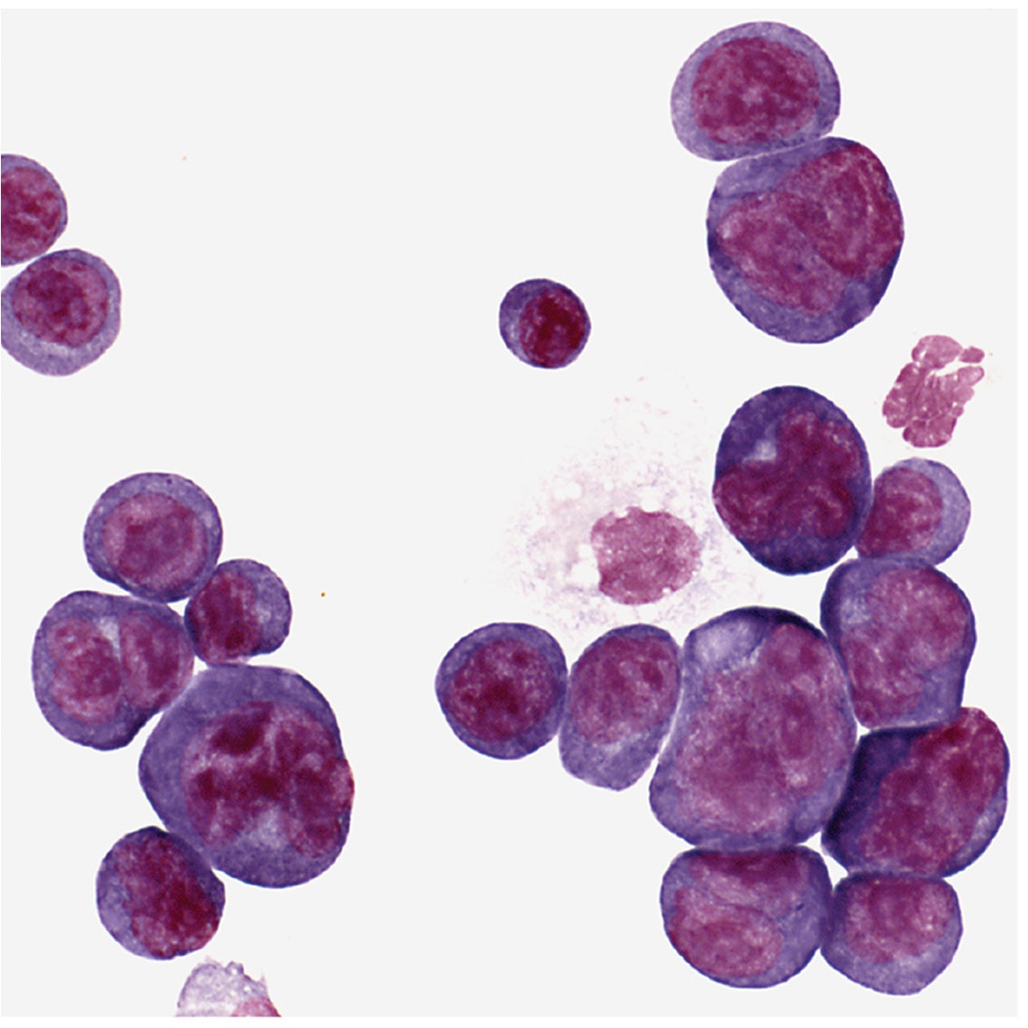
Tumor cells
| Span | ||
|---|---|---|
| ||
| National Library of Medicine |
Potter was usually at his laboratory bench using this microscope. Shown are plasma cells with darkly stained nuclei. The clear spots next to the nucleus are perinuclear “hoffs” and are filled with newly synthesized protein, in this case, antibody.
| Span | ||
|---|---|---|
| ||
| Donated by Dr. Beverly Mock |
| Span | ||
|---|---|---|
| ||
| Donated by Dr. Beverly Mock |
| Span | ||
|---|---|---|
| ||
| Donated by Dr. Beverly Mock |
| Span | ||
|---|---|---|
| ||
| Donated by Dr. Beverly Mock |
| Button | ||||||
|---|---|---|---|---|---|---|
|
Overview
Content Tools
ThemeBuilder