
Onsite Exhibits Gallery
Please visit the Exhibit Maps page for more information about the physical location of the exhibits listed on this page.
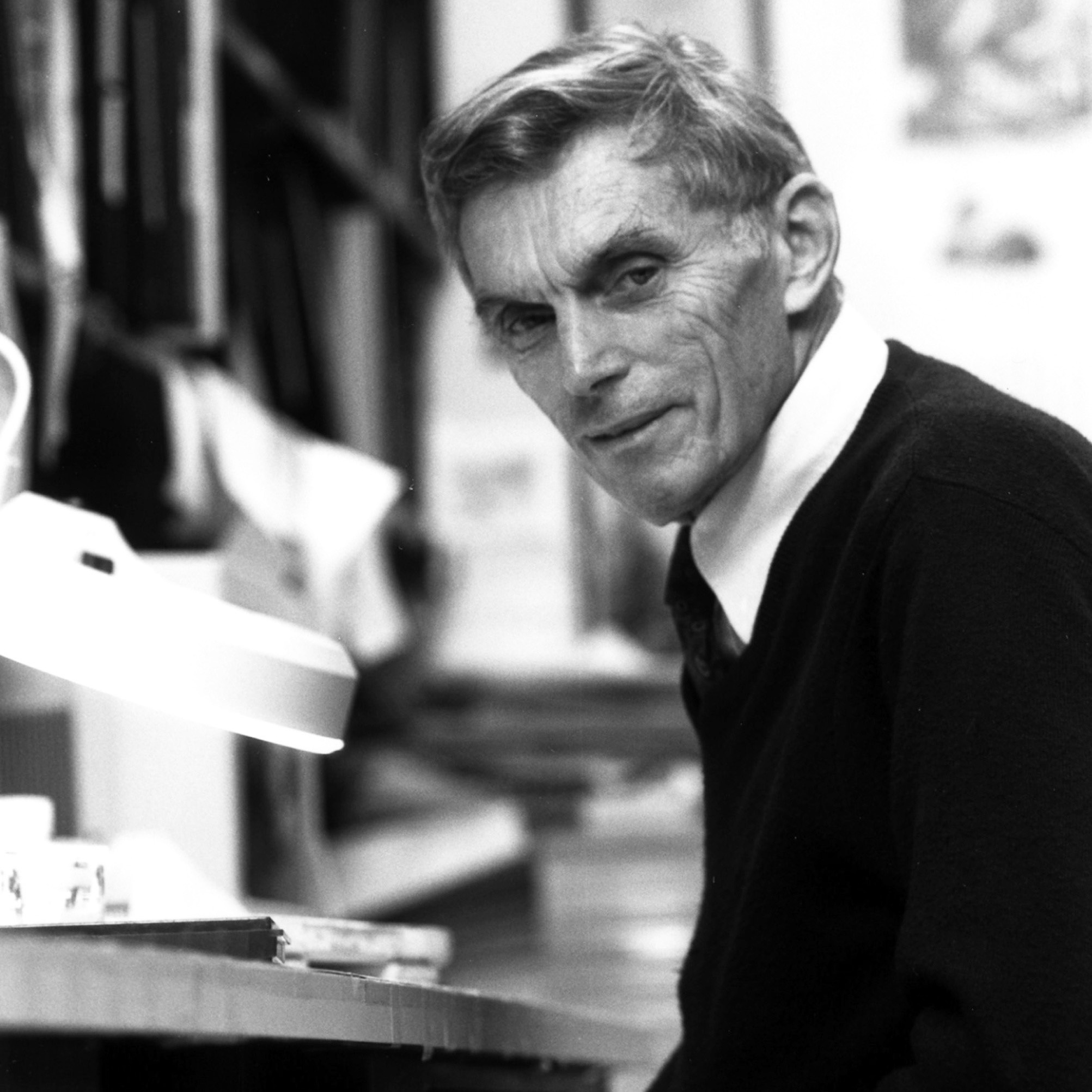
How are proteins made? How do they fold, and what role does structure play in their function? Chris Anfinsen's investigations answered these questions; they also led to a Nobel Prize.
Michael Potter investigated the twin questions of what causes cancer and how we produce the antibodies called immunoglobulins which protect us from disease.

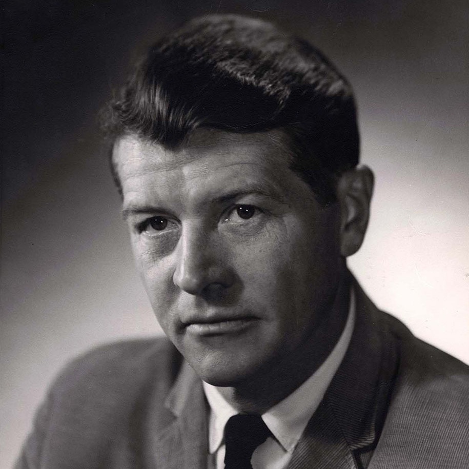
Explore the Nobel Prize-winning work of Marshall Nirenberg, who deciphered the genetic code with the help of NIH colleagues, enabling genetics to become a central scientific field.
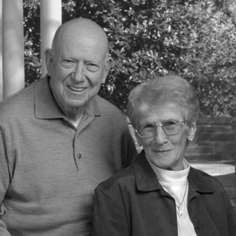
The scientific power couple of Thressa and Earl Stadtman developed a unique way to train scientists; they each made significant scientific contributions too.
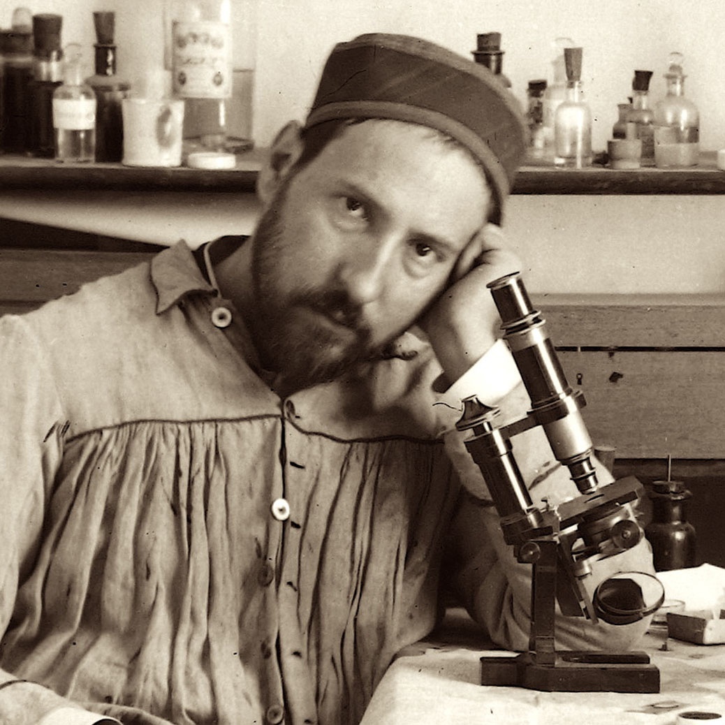 Learn about the first person to describe the nervous system, including intricate neurons, in exquisite and artistic detail was Santiago Ramón y Cajal.
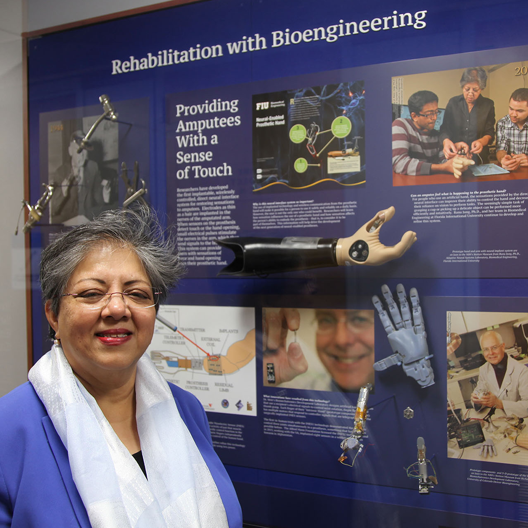
Learn about cutting-edge research funded by the National Institute of Biomedical Imaging and Bioengineering.
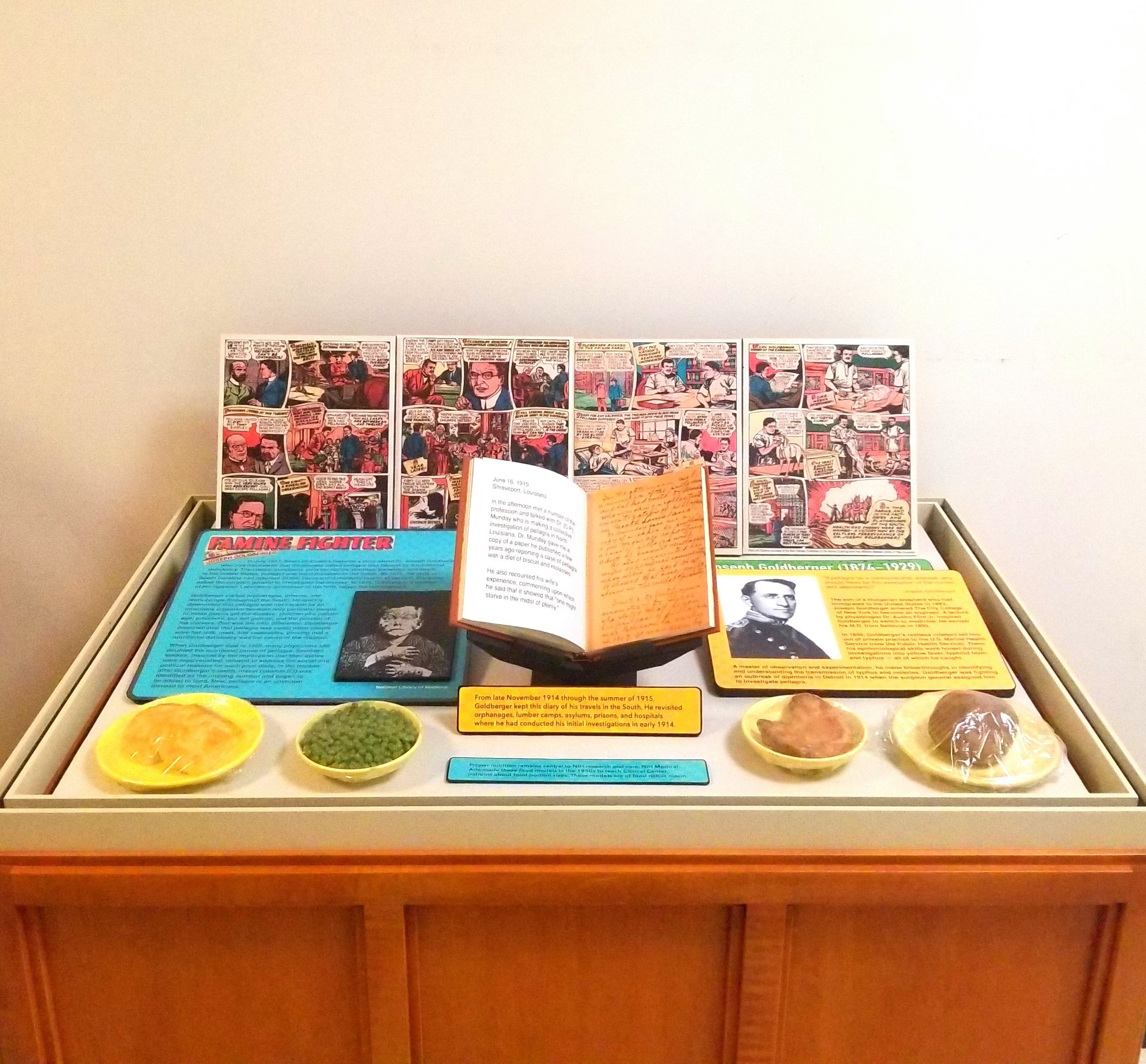
Dr. Joseph Goldberger discovered the cause of pellagra, a disease that killed many poor Southerners in the early part of the 20th century. His finding that pellagra was caused by a diet deficient in vitamin B was met by political and social resistance.
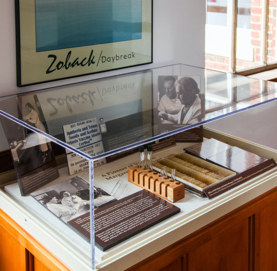
Margaret Pittman
Margaret Pittman arrived at NIH in 1936, beginning a career that would span 57 years and make her an internationally renowned expert on vaccines and serums, as well as the first female laboratory chief at the NIH.
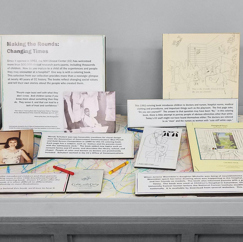
Changing Times
Who would think that coloring books would provide a glimpse at nearly 40 years of Clinical Center history, each reflecting changing times and telling their own stories about the people who created them?
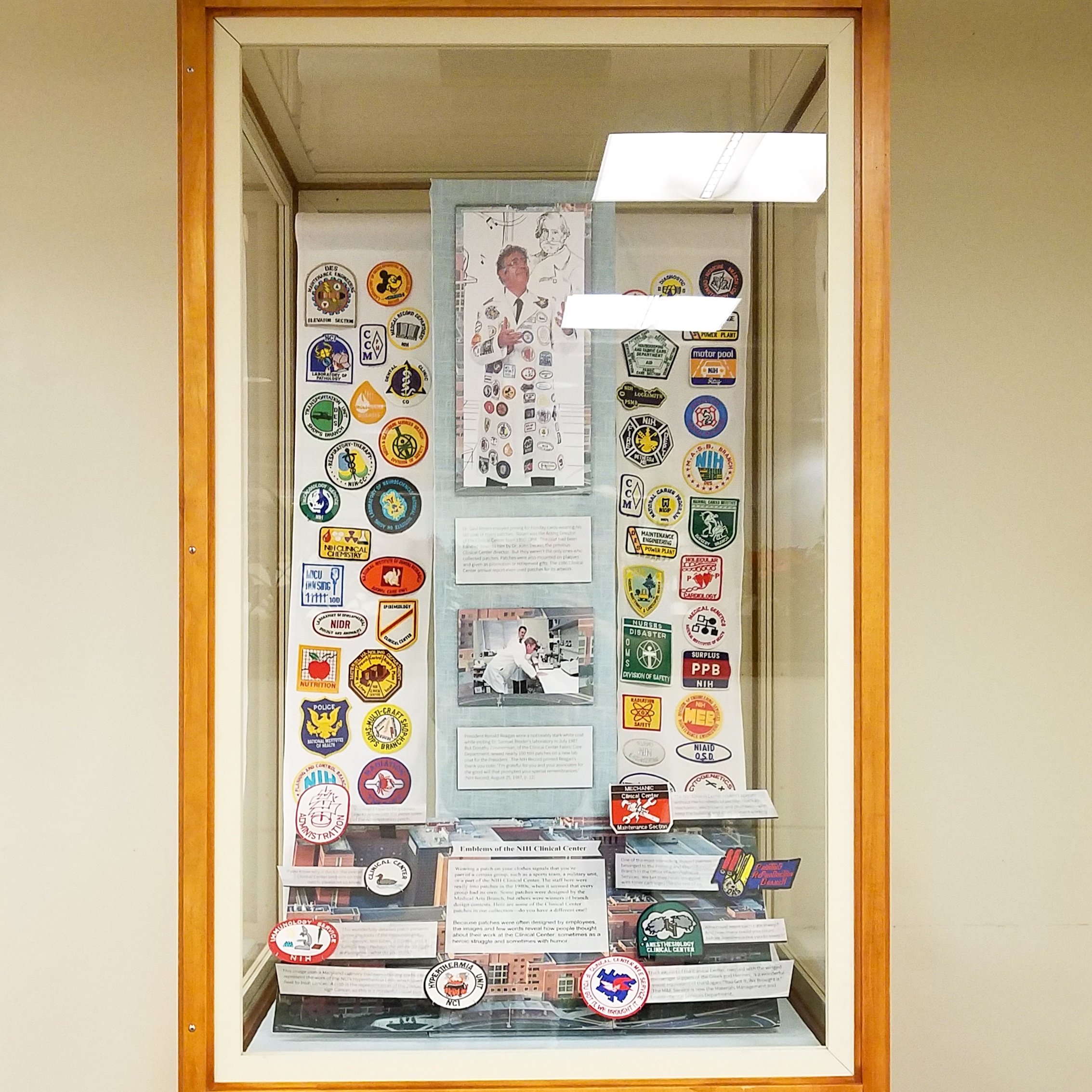
Pretty Patches
Because employees designed these patches, they reveal how people thought about their work at the Clinical Center—sometimes as a heroic struggle and sometimes with humor.
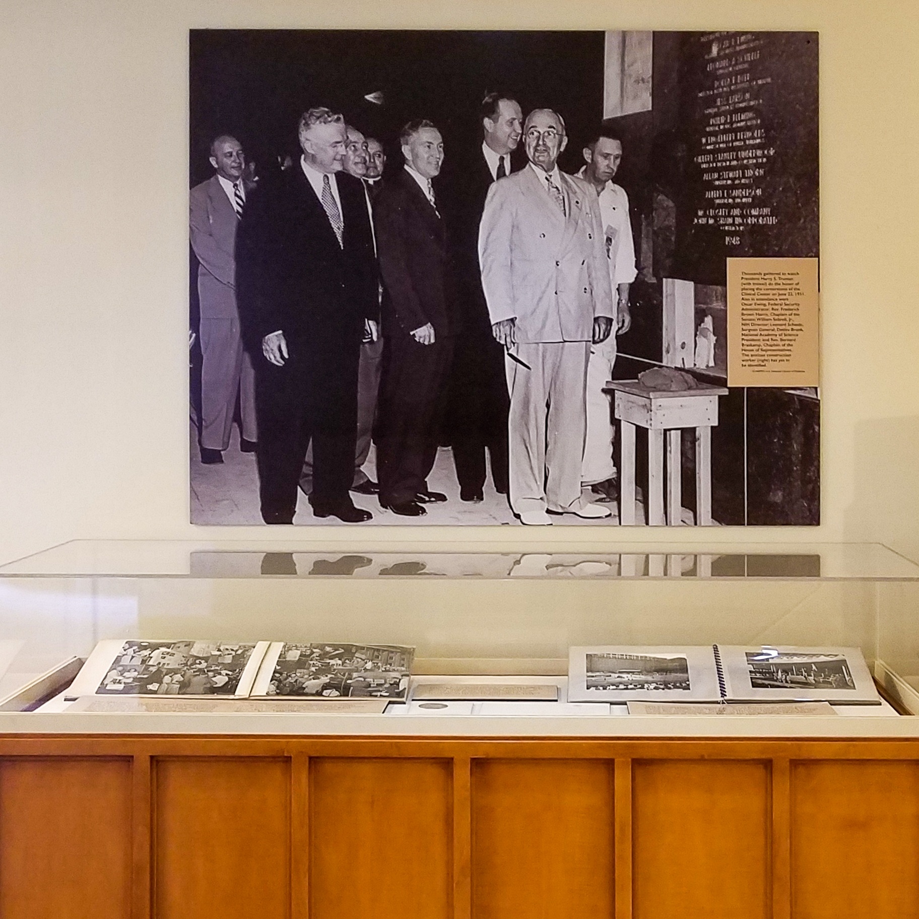
Harry Truman
See photo albums from the 1948 Open House at NIH, which helped explain the Clinical Center concept to the public, and President Harry Truman's laying of the hospital's cornerstone in 1951.
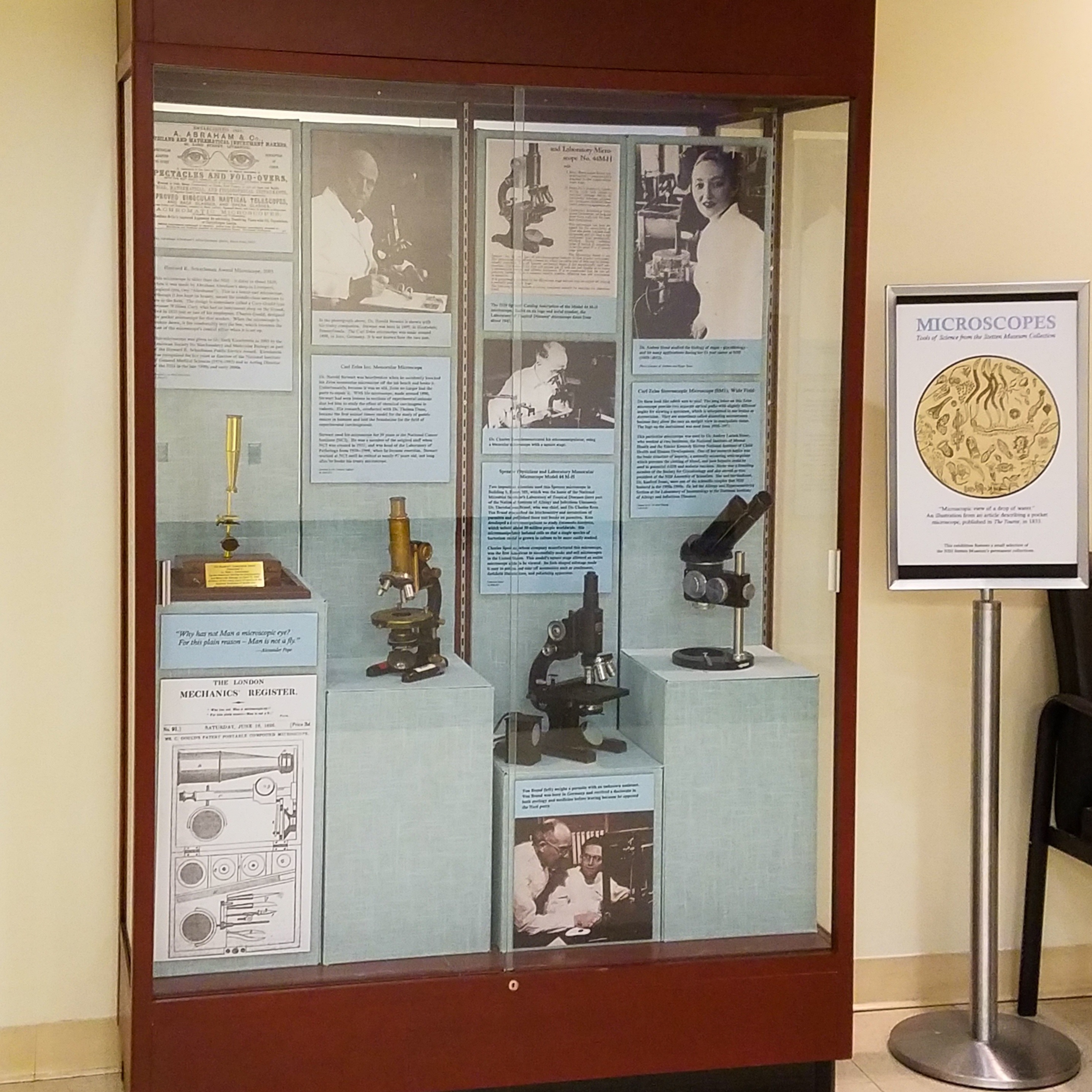
Microscopes
Learn about the scientists behind their microscopes and the vast array of microscopes used at the NIH.

Siemens 1-A Electron Microscope
All sorts of viruses were visualized for the first time on this Siemens 1-A Electron Microscope used by Albert Kapikian.
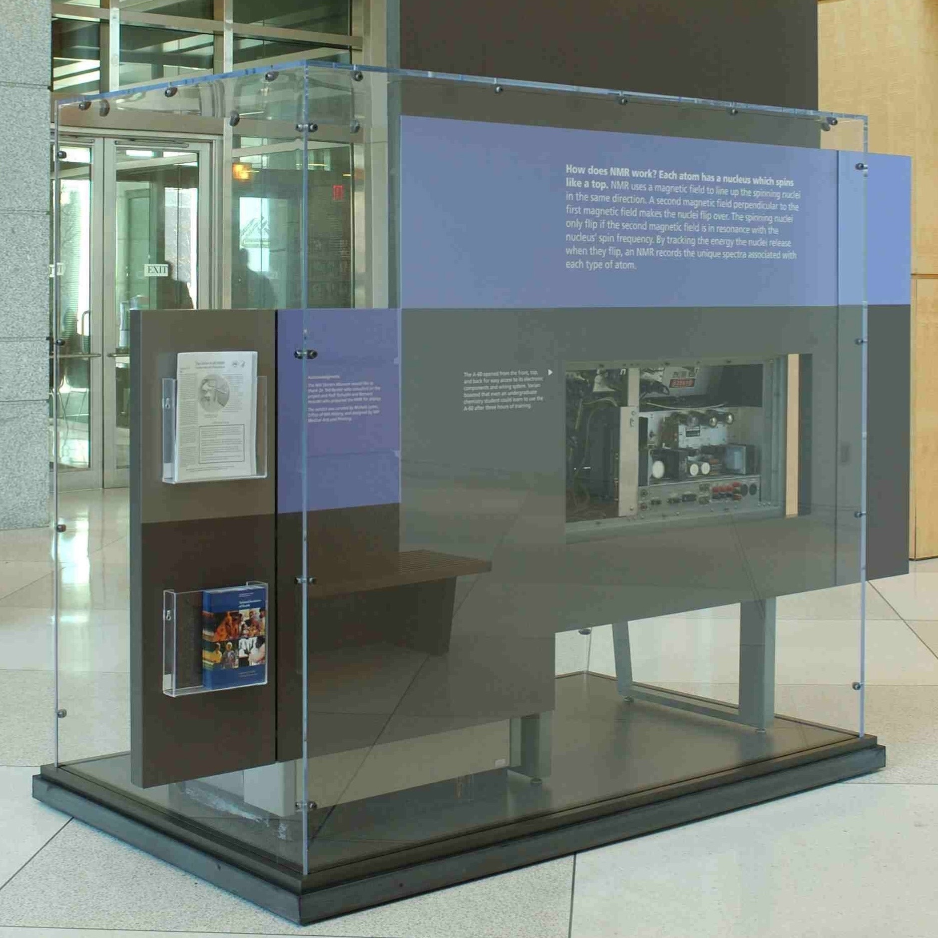
Varian A-60 NMR
The Varian A-60 NMR (nuclear magnetic resonance) spectrometer was the first low-cost instrument of its kind, producing a magnetic resonance image (MRI) that NIH scientists used to study topics such as how the brain develops as children grow.
Overview
Content Tools
ThemeBuilder