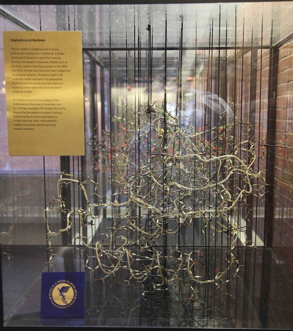
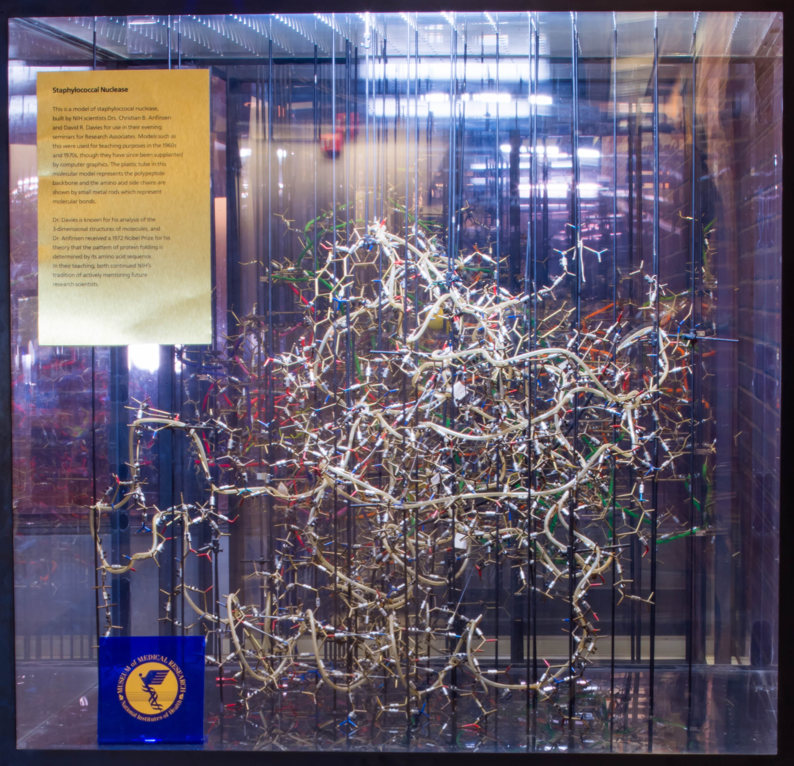
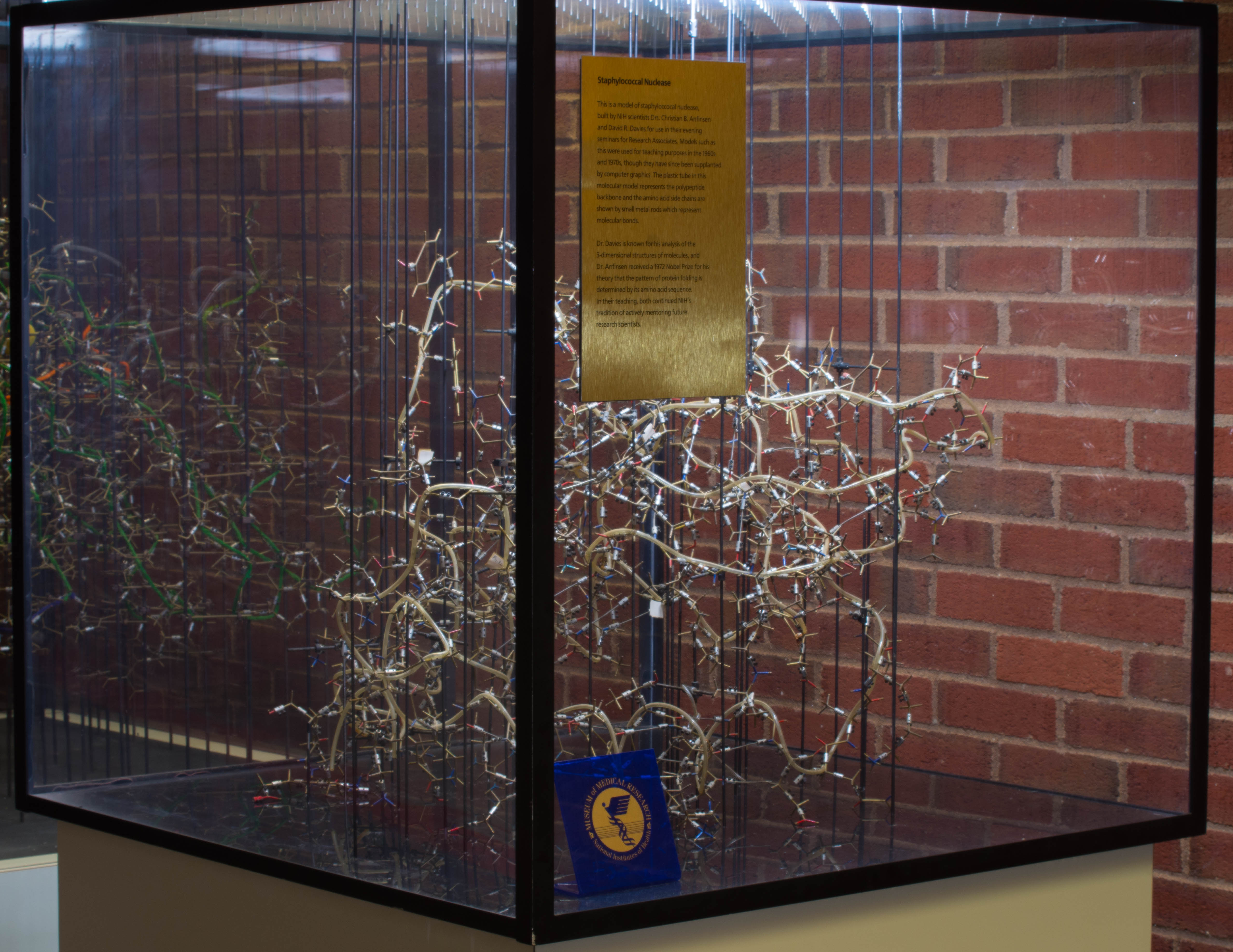
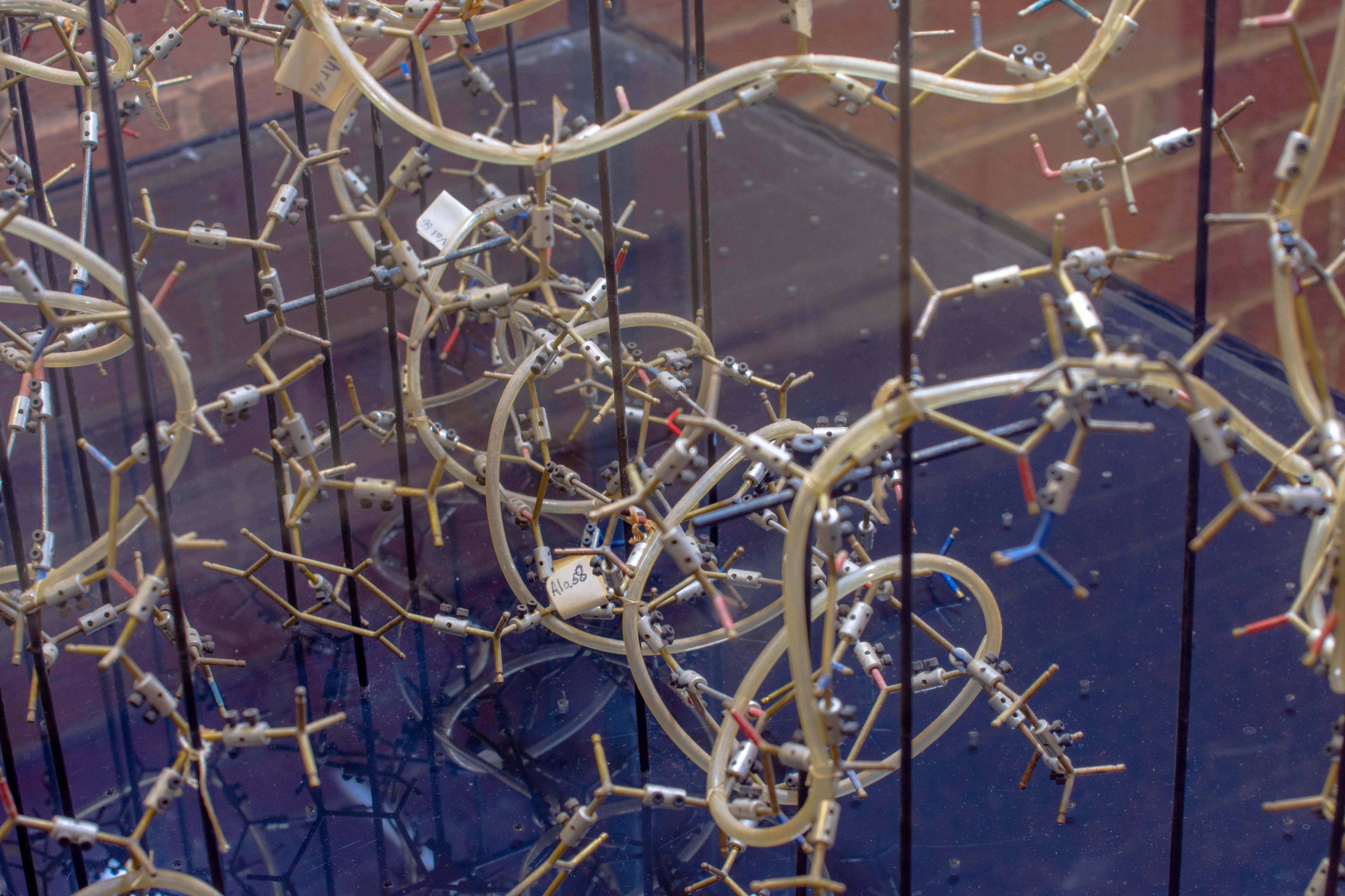
Staphylococcal Nuclease Model, c. 1965
Donated by Dr. Alan Schechter, Accession 89.0001.380
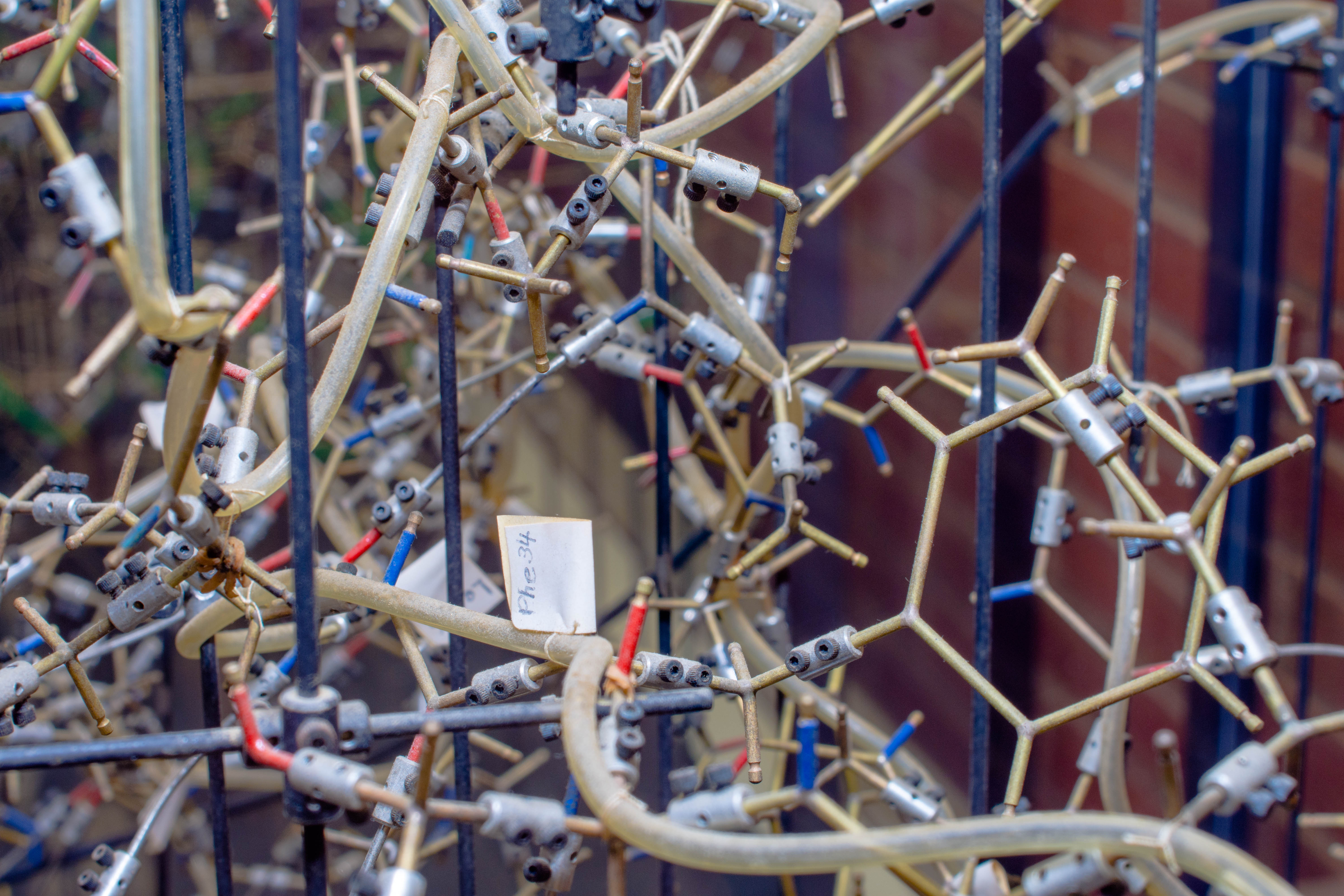
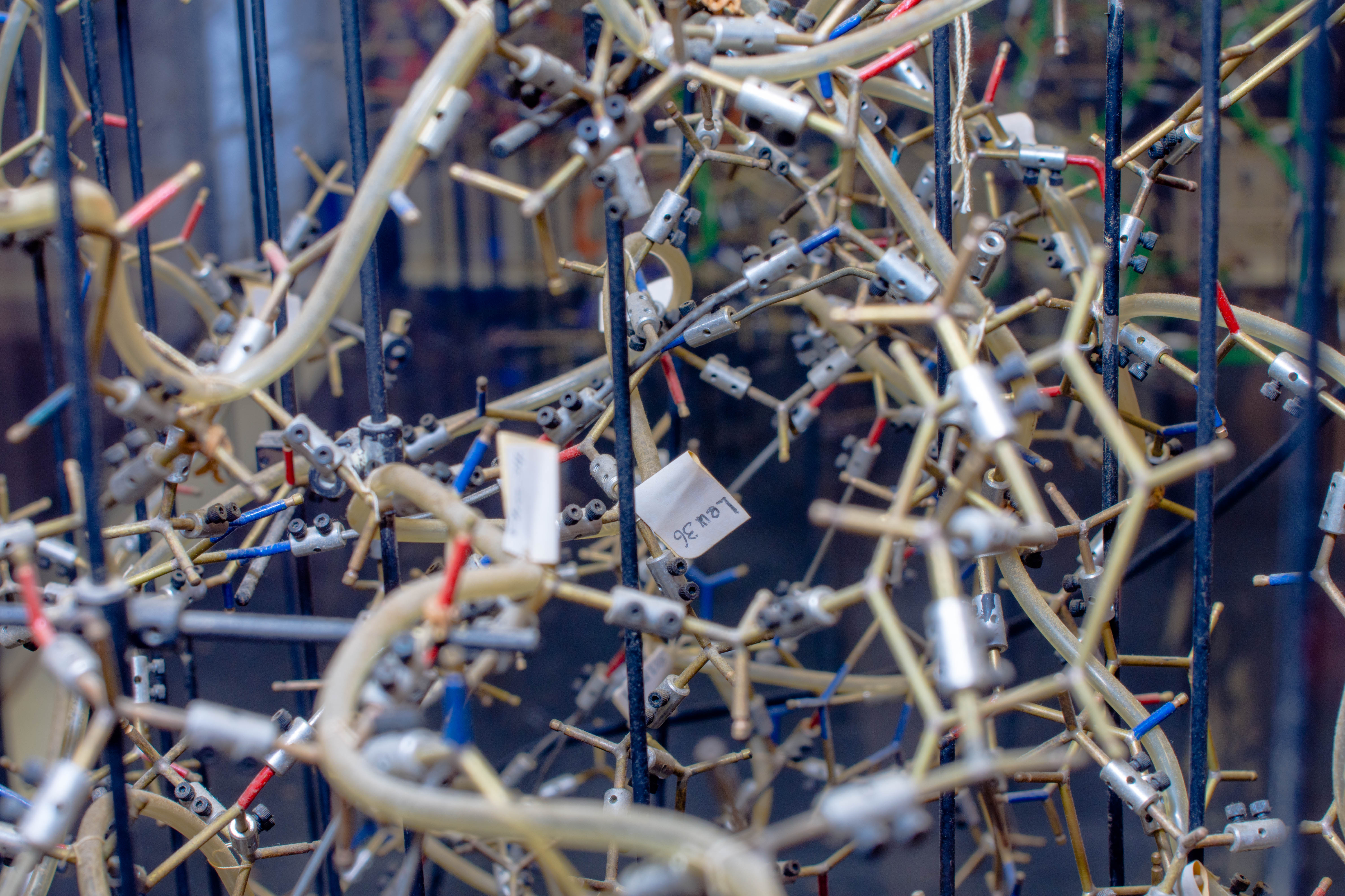
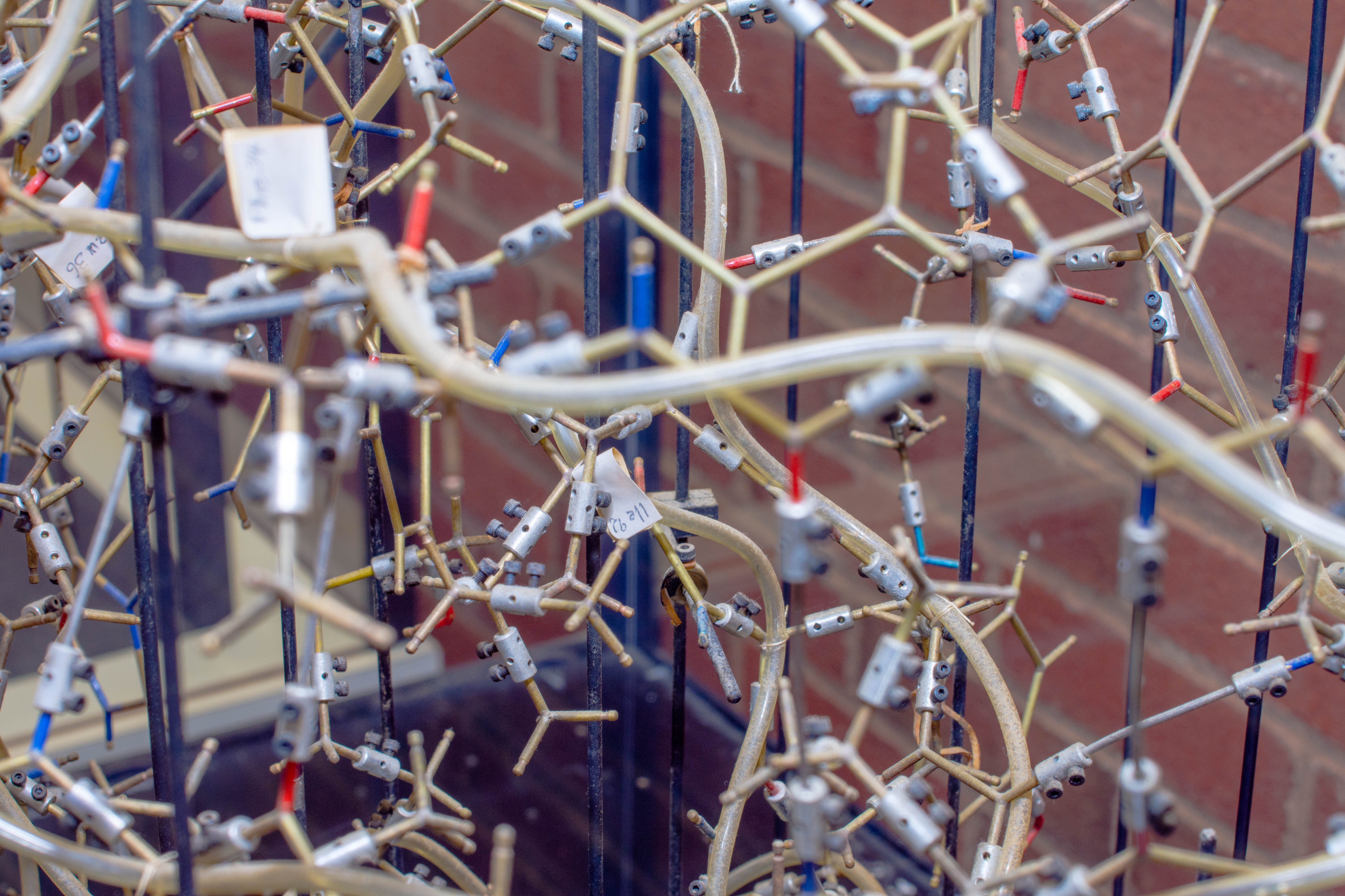
Staphylococcal nuclease was the focus of extensive experimental work under Dr. Christian Anfinsen from about 1963 to 1975. This molecular model was built as part of the educational program of Protein Chemistry evening seminars for Research Associates conducted throughout the 1960’s by Drs. Anfinsen and David Davies. It’s based on the x-ray crystallographic analysis done at the Massachusetts Institute of Technology under the direction of Professor Albert Cotton. The polypeptide backbone (traced by the plastic tube) and the amino-acid side chains are shown by small metal rods which represent molecular bonds in these models.
Donated by Dr. Christian Anfinsen
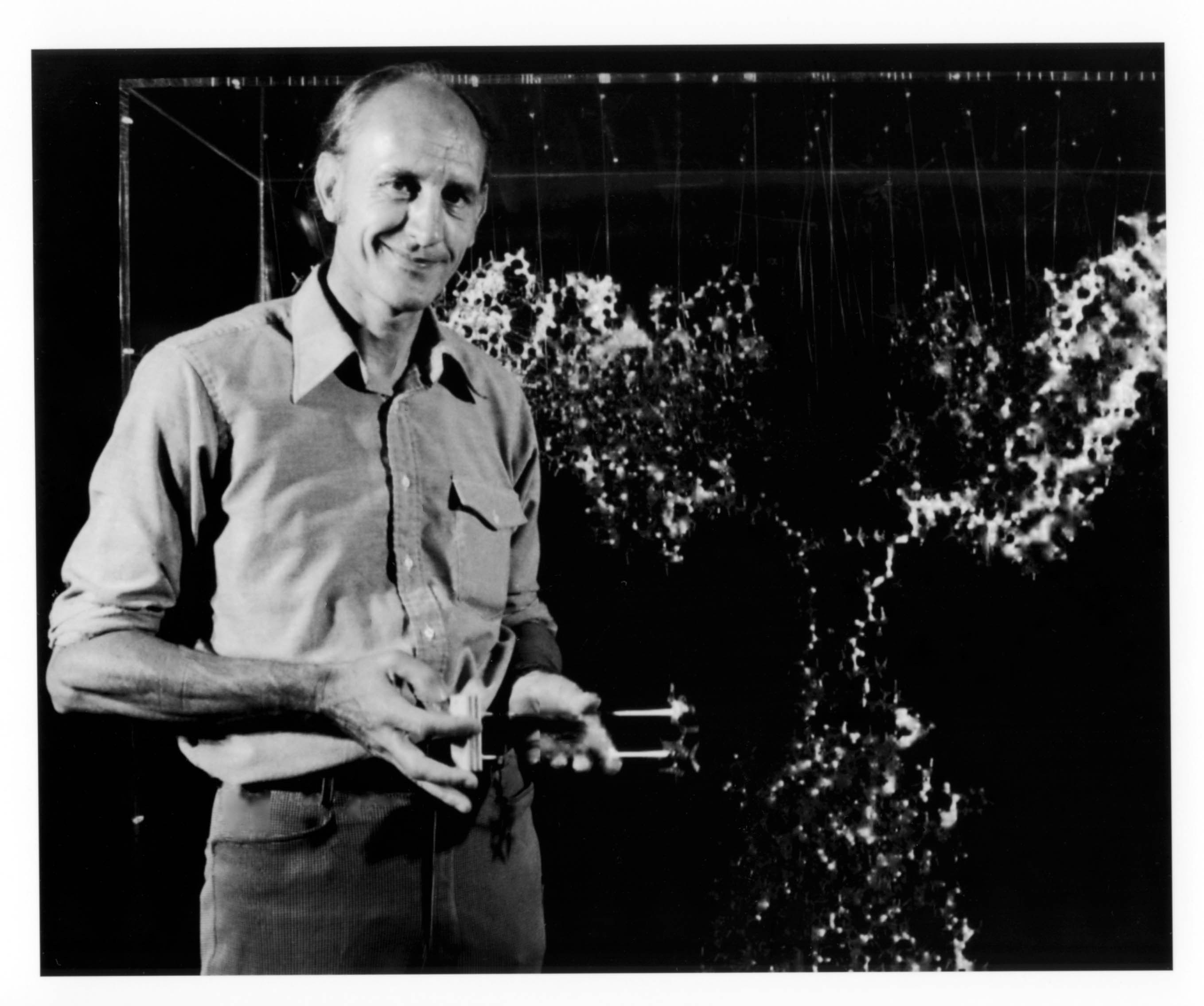
David Davies had studied under John Kendrew at Cambridge in England as a visiting scientist from the National Institute of Mental and so was uniquely trained to construct such models.
In 1958, Kendrew had reported the first visualization of a protein’s structure when using low resolution x-ray crystallographic analysis on the protein myoglobin. A model of the protein had to be built for publication purposes. In 1965, Kendrew approached A. A. Barker, an employee of the Cambridge University Engineering Laboratories, asking him to make more models for him. Barker used a ball and stick type of model so that the center was open to be visible and adjustable. Putting together the models was made easier when A. Beevers, a professor of chemistry at the University of Edinburgh, invented a machine to drill holes in small plastic balls. The ball and stick set-up for the protein models became known as Kendrew components.
Donated by Dr. Christian Anfinsen
Dr. David R. Davies graduated from Oxford University in 1949 and received a Ph.D. in 1952. In 1955, he joined NIMH and moved to NIDDK six years later. His research used x-ray crystallography to determine the three-dimensional structure of proteins and nucleic acids.
Read our oral histories with David Davies
- https://history.nih.gov/archives/downloads/davidrdaviestranscript121099.pdf
- https://history.nih.gov/archives/downloads/davidrdaviestranscript112299.pdf
Additional Images
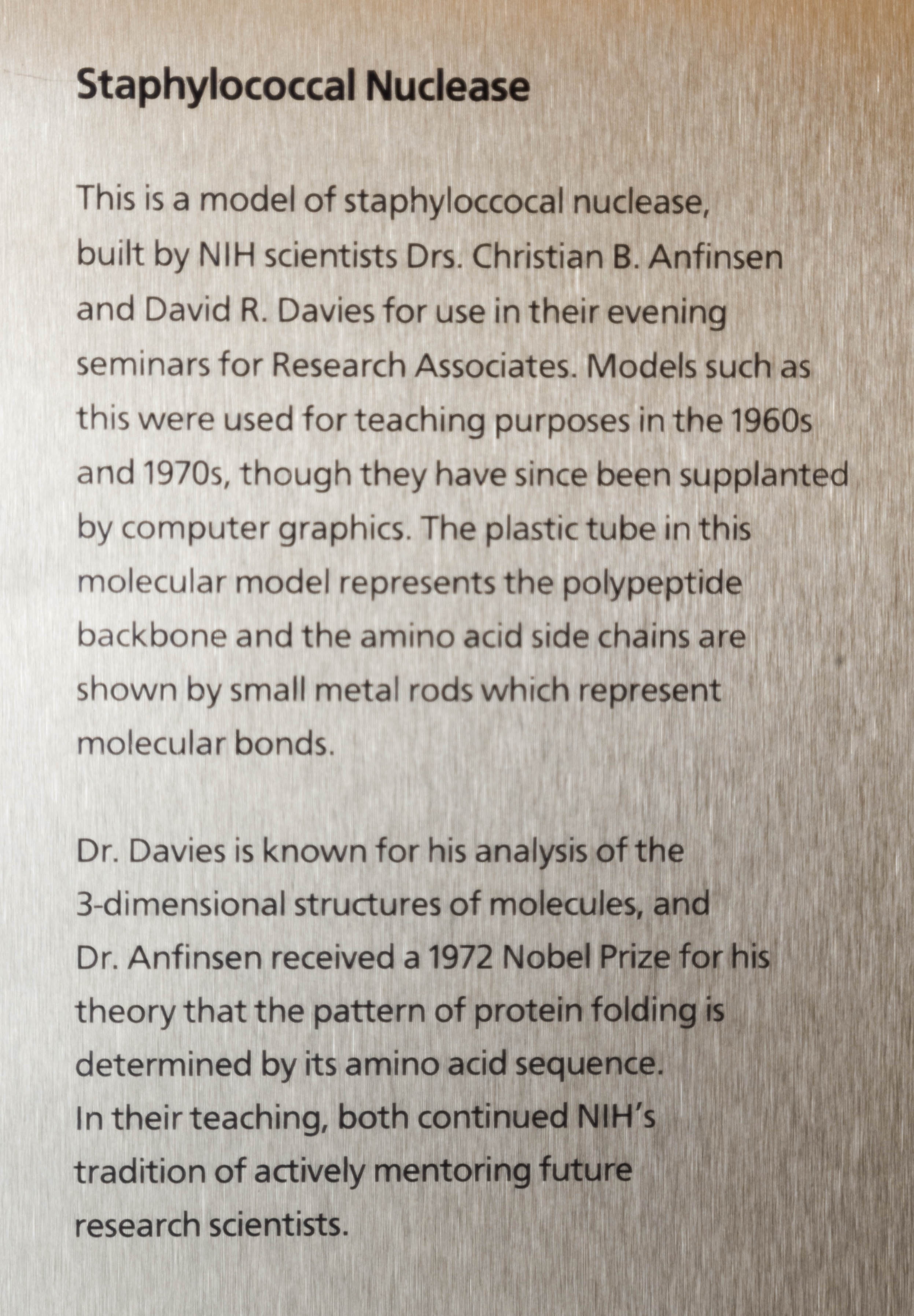
Staphylococcal Nuclease This is a model of staphylococcal nuclease built by NIH scientists Drs. Christian B. Anfinsen and David R. Davies for use in their evening seminars for Research Associates. Models such as this were used for teaching purposes in the 1960s and 1970s, though they have since been supplanted by computer graphics. The plastic tube in this molecular model represents the polypeptide backbone and the amino acid side chains are shown by small metal rods which represent molecular bonds. Dr. Davies is known for his analysis of the 3-dimensional structures of molecules, and Dr. Anfinsen received a 1972 Nobel Prize for his theory that the pattern of protein folding is determined by its amino acid sequence. In their teaching, both continued NIH's tradition of actively mentoring future research scientists.
3d models such as this have largely been supplanted by 3d graphics and resources such as the RCSB Protein Data Bank website: Staphylococcal Nuclease interactive graphic.


