
The Canyon Creek Schoolhouse Laboratory’s Mission: Rocky Mountain Spotted Fever
When researching a disease, scientists and physicians ask some pretty basic questions: Is there a disease? What causes it? How do you get it? How do you know you have that specific disease? What does it do to you? Can we treat or cure it? Can we prevent you from getting it?
When the Canyon Creek Schoolhouse laboratory opened in 1921 to investigate Rocky Mountain spotted fever (RMSF), only one of these questions had been answered definitively: Is there a disease? Yes, definitely! Other questions had partial answers that had created more questions. Two big unknowns were how to treat or cure RMSF and how to prevent people from getting it in the first place. The mission of the laboratory was to answer as many of these questions as possible.
What causes RMSF?
Collecting Ticks
Although it was known that RMSF was caused when the Rickettsia rickettsii bacteria infected a Dermacentor andersoni tick that then bit a human, more questions remained. Was the bacteria spread by other species of ticks? And exactly how did the ticks get infected? To find out where the ticks were picking up the infection and to answer a number of other questions, ticks had to be collected in the wild.

A staff member of the Canyon Creek Schoolhouse Laboratory—possibly Dr. Ralph Parker, C.M. Salisbury, or George Cowan—dragged a white flannel flag over brush and grass to gather ticks in the Bitterroot Valley, Montana. He wore a white jump suit over his regular clothes, tucking his pant legs into the top of his high, laced boots. When they returned from such outings, the men would check each other closely in case a tick had attached itself to one of them.
Image: Office of NIH History and Stetten Museum, 1522

In 1923, a mountain goat (not this mountain goat kid) taken by George Cowan had over 1,000 ticks engorged ticks on it. It was with these ticks that Dr. Roscoe Spencer came up with his idea for an RMSF vaccine.
Image: Montana Memory, 264
Cataloguing Ticks
As the field researchers gathered many species of ticks and ticks in many stages of their life cycle, Dr. Robert Cooley developed a huge collection of them. Cooley was the head entomologist at Canyon Creek Schoolhouse laboratory. This collection would help later researchers solve questions about other tick-borne diseases.

Dr. Robert Cooley shows off his world-class tick collection. The second picture was taken in Building One (which opened in 1928), not the Canyon Creek Schoolhouse laboratory, on December 3, 1946. Cooley had been developing this collection since his first days studying RMSF forty years before.
[RML may have a photo like these in high res that could be used instead.]
Image: Wikimedia Commons
How do you get RMSF?
Occurrence
One way of finding out how a disease is spread is by looking at where it occurs. Rocky Mountain spotted fever came to be studied in Montana’s Bitterroot Valley because the bites of the western slope’s ticks caused a particularly deadly infection, meaning that more people died. By mapping and charting where they found ticks or where people got RMSF, the researchers could find the ticks and animals they needed to study. Mapping turned up interesting insights, such as one area may have a heavily infected tick population, but an area across a stream did not. Ticks do not like to swim.
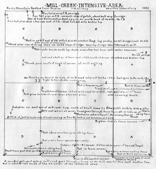
C.M. Salisbury drew this grid map of an area of land near Mill Creek in 1922. Included were the sites of stumps, trees, and roads. The researchers could use it to keep track of where they collected ticks.
Image: Montana Memory 961b -WE HAVE OTHER MAPS TO SCAN TO USE
Life cycle of ticks
Studying the life cycle of ticks provided important pieces of the RMSF puzzle. Knowing the life cycle meant that scientists could study questions such as: When did the ticks pick up the bacteria causing RMSF—was it when they were newly hatched? When they were adults? And was the infection passed on to a new generation of ticks through the eggs? The answers to these questions could be used in control efforts and in developing a new vaccine.

The life cycle of the tick: a fertilized female lays thousands of eggs on the ground. When they hatch as larvae, they attach to small animals and feed. Then they drop off to digest their meal, and molt (shed their old shell) to become nymphs. The first winter they spend as unfed nymphs.
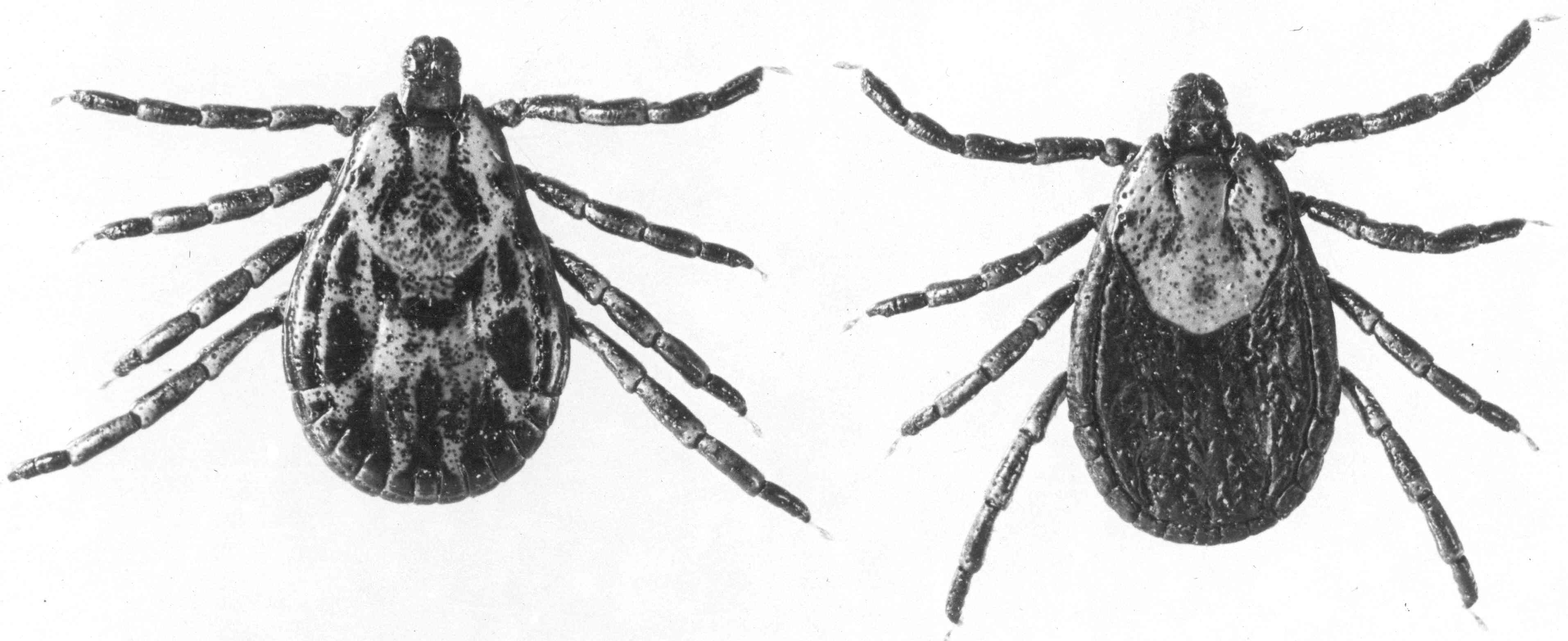
In spring, the nymphs once again attach to an animal and feed, drop to the ground, and digest their meals. This time they molt to adults, like these Rocky Mountain wood ticks, Dermacentor andersoni. They spend their second winter as unfed adults.

The next spring they climb grass and bushes to attach to animals.

But as adults, ticks mate as well as feed. This is when the ticks can get the Rickettsia rickettsii bacteria from the animals they feed on. Even though the bacteria don’t hurt the ticks, it infects all of their cells, and is passed on through the egg. The next generation of ticks will be infected.
Images: Office of NIH History and Stetten Museum, 1454-58
Environmental studies
To understand the entire picture of RMSF transmission, the entomologists at the Canyon Creek Schoolhouse laboratory began environmental studies of the relationships between the distribution of vegetation, rodents, ticks, and humans.

“Valuable Experiment: Please Do Not Disturb” reads a sign put up by one of the RMSF researchers in one area undergoing an ecological study.
Image: Office of NIH History and Stetten Museum, 1479
Constant collecting
As RMSF gave up its mysteries, researchers still had other tick-borne diseases to study. They constantly collected ticks where outbreaks of Rickettsial diseases occurred.

In 1929, LeRoy Jones, Harley G. Sargent, Harry L. Sargent, and James Kerlee posed at the top of Blodgett Canyon in the Bitterroot Range. Each holds several white cloth bundles tied to a stick for specimen collection. James Kerlee’s brother, Arthur LeRoy Kerlee, had died the year before of RMSF that he had acquired in the laboratory.
Image: Office of NIH History and Stetten Museum, 1449
How do you know you have RMSF?
Diagnostic test
In a world filled with ticks and the diseases they carry—and diseases that have similar symptoms—being able to diagnose a disease quickly might be beneficial for treatment or for clues to how to control it. The classic feature of RMSF, the rash, appears days after the initial infection. The other symptoms can be confused with diseases such as typhus.
In 1916, the Weil-Felix reaction was developed to diagnose epidemic typhus. In 1928, LeRoy Kerlee and Dr. Roscoe Spencer did their own tests on guinea pigs, rabbits, and people at the Canyon Creek Schoolhouse laboratory. They reported that the Weil-Felix reaction could diagnose RMSF. The test became widely used to diagnose many Rickettsial diseases; although it has been replaced by newer diagnostic techniques such as indirect immunofluorescence, it can be useful in parts of the world without access to such technology.
Kerlee and Spencer’s results were published shortly after Kerlee died of Rocky Mountain spotted fever in 1928. Learn more about Kerlee [link to his bio].
“Rocky Mountain Spotted Fever: A Preliminary Report on the Weil-Felix Reaction,” A. L. Kerlee and R. R. Spencer, Public Health Reports (1896-1970), Vol. 44, No. 4 (Jan. 25, 1929), pp. 179-182
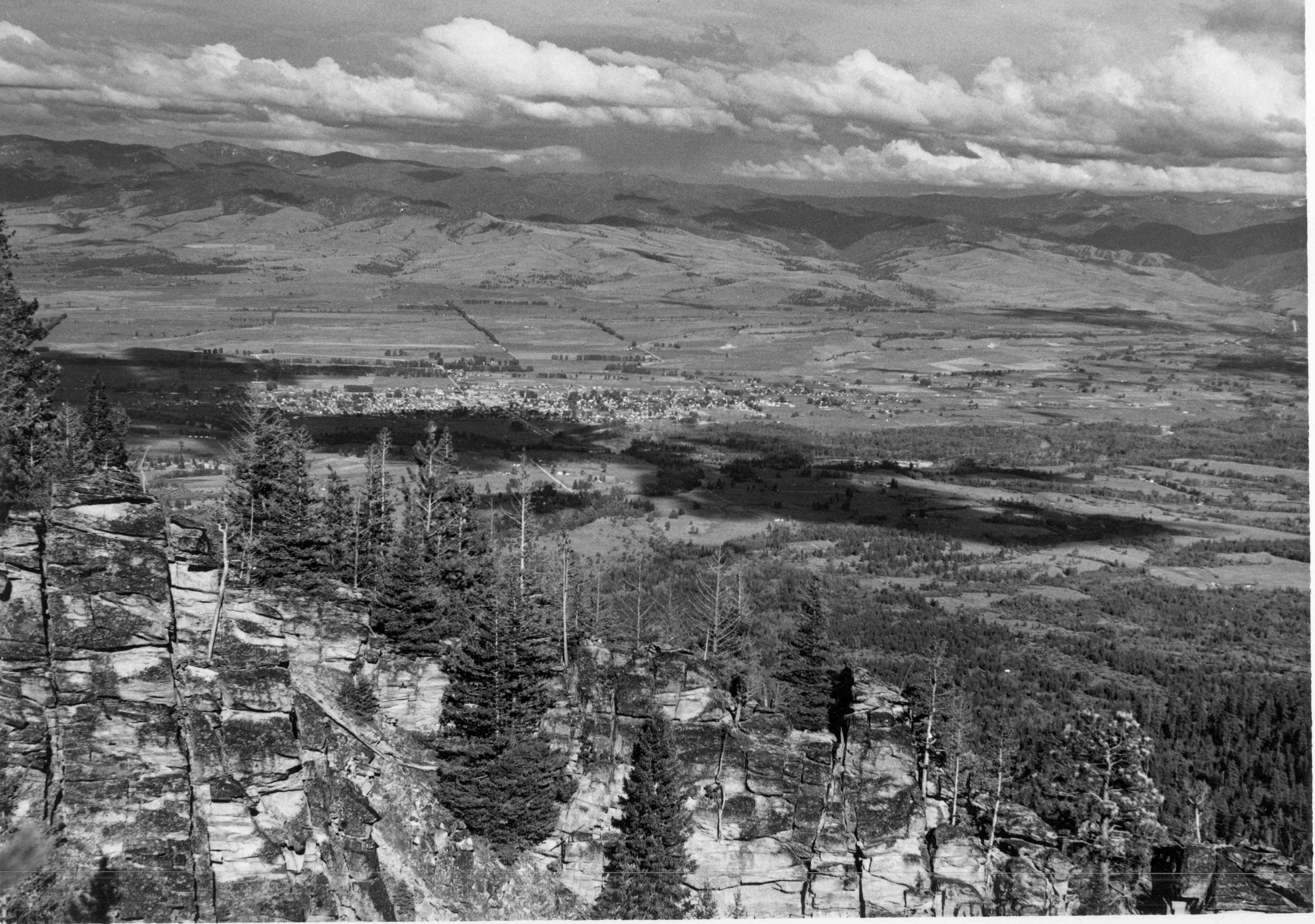
Overlooking Hamilton, Montana, where the Canyon Creek Schoolhouse laboratory was located. How many ticks were in that valley?
Image: Office of NIH History and Stetten Museum, 1562-3
Can we prevent RMSF?
The best way to deal with a deadly disease is to not get sick with it. But avoiding a disease can depend on how it’s spread, where you live or what you do for a living, and the possibility for a protective vaccine.
Control efforts
Because it was known that ticks carried RMSF, one way to eradicate the disease was to keep people from getting bitten by infected ticks. That could mean changing the way that ranchers, shepherds, woodsmen, and others did their jobs. Or it could mean eradicating the animal hosts of the tick in an area of heavy infection. Both methods were tried.

To protect the workers most in contact with animals that could harbor ticks, Dr. Robert Cooley, the Montana State Entomologist and head Entomologist at the Canyon Creek Schoolhouse laboratory, recommended dipping livestock in disinfectants to kill the ticks, as was being done in this photo.
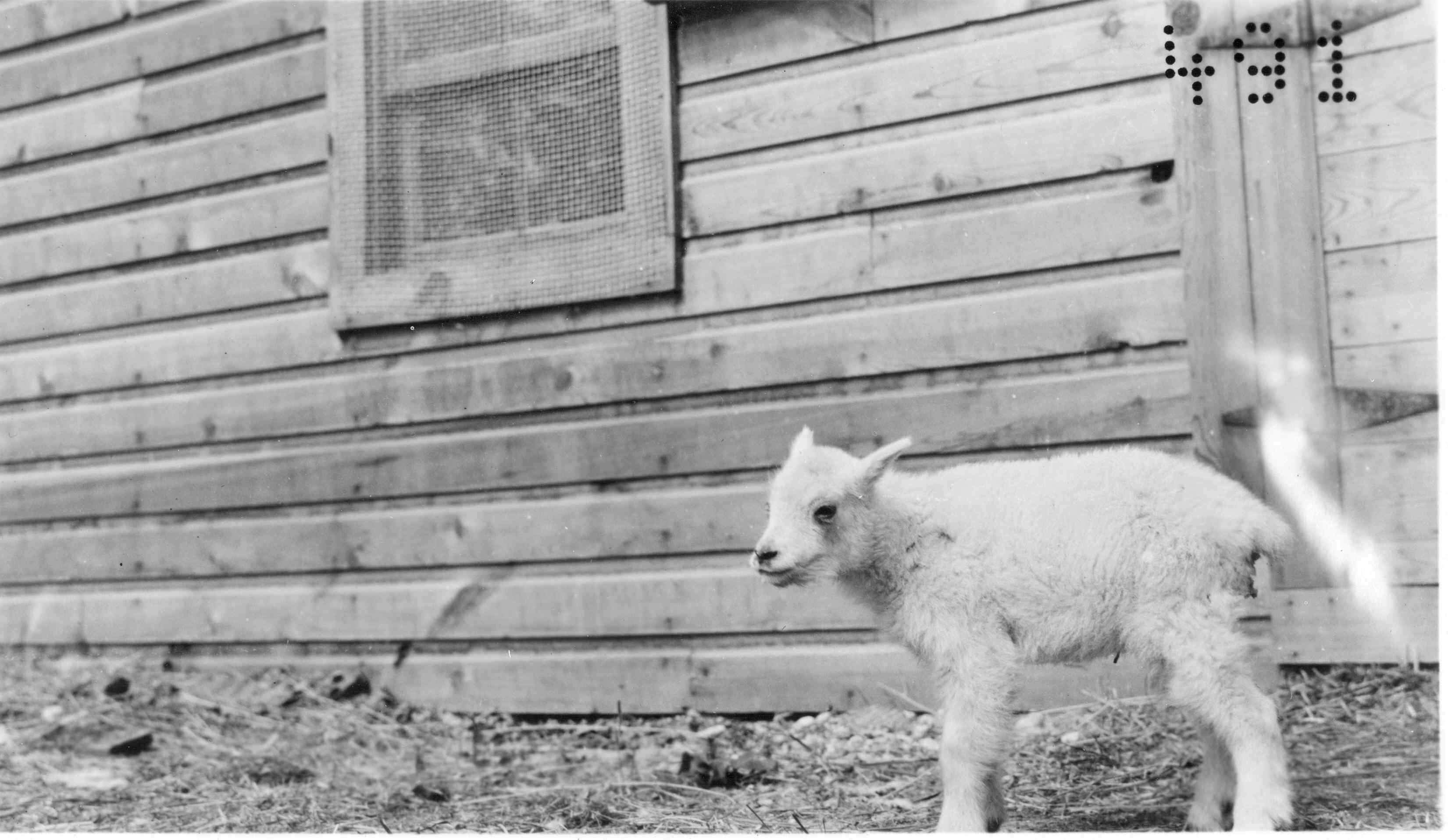
There was another approach to tick control: eradicating the rodents and other small animals in an area where RMSF outbreaks occurred, often by using poison. This mountain goat kid at the Canyon Creek Schoolhouse laboratory represents just one kind of animal that can carry the bacteria which infects ticks, which then infect humans. The bid to eradicate small animals in areas infested with RMSF failed; it wasn’t clear which animal or animals gave the infections to ticks, and new animals kept moving in.
Images: Office of NIH History and Stetten Museum, 1520 and 1469
Vaccine
One of the surest ways to stop the spread of a disease is to develop a vaccine against it. At the Canyon Creek Schoolhouse laboratory, bacteriologists (Dr. Roscoe Spencer) and entomologists (Dr. Ralph Parker) worked together to that end. Despite the limited technology and understanding of bacteriology of the 1920s, once Spencer and Parker began to work together in 1921, a vaccine was developed in less than three years.

As illustrated by this photo of two RMSF researchers, the development of a RMSF vaccine was only possible because of cooperation between state and federal agencies, scientific disciplines, and the research staff.
Image: Office of NIH History and Stetten Museum, 1114
Inventing the Vaccine
Dr. Roscoe Spencer came to the Canyon Creek Schoolhouse laboratory from Washington, D.C. in spring 1921, when the ticks were out. After the laboratory’s field workers collected ticks, Spencer would test the ticks for RMSF by taping them to a guinea pig; if the guinea pig came down with RMSF, the ticks were infected. But this process was messy and dangerous and slow, so he decided to grind up the ticks and inject them under the skin of the guinea pigs. Still, none of them got sick. Then he took samples from guinea pigs that were sick with RMSF. He injected the samples into guinea pigs that had been injected with ground-up ticks and into those guinea pigs that had not. The guinea pigs that had previously been injected with the ground-up ticks did not get sick.
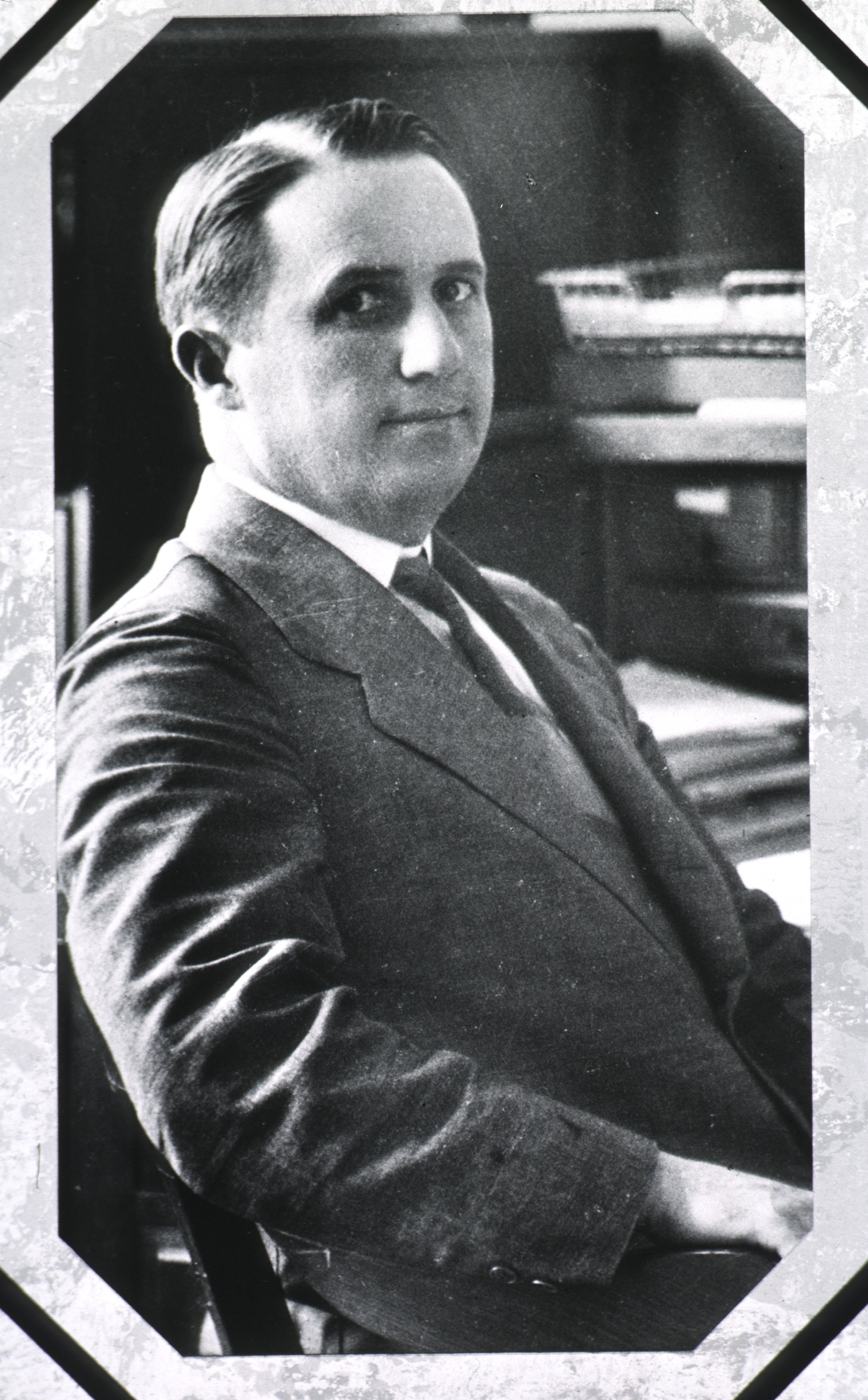
Dr. Roscoe Roy Spencer.
Image: National Library of Medicine, 101429481
One day field worker George Cowan brought in a mountain goat covered with nearly a thousand engorged ticks. The researchers plucked the ticks off and decided to see what would happen when they ground them up and injected them into healthy guinea pigs. Every guinea pig got sick and died. The control group were guinea pigs that had been injected with ground-up unfed ticks; they did not get sick.
To be sure of the results, Spencer took some unfed ticks, attached them to a sick guinea pig to feed, ground up the ticks, and injected them into healthy guinea pigs. They all died. As Lucy Salamanca dramatically wrote: “They had proved it was the meal of blood that had turned a harmless tick into an agent of death!”
(Quote from “Tick Conquered,” Lucy Salamanca, Washington Evening Star, March 28, 1937, page F-4.)

Ticks feeding
Image: Office of NIH History and Stetten Museum, 1465-3
Dr. Ralph Parker and his group at the Canyon Creek Schoolhouse laboratory became responsible for raising ticks and guinea pigs to feed them. The guinea pigs lived in cages swathed with white cotton as shown in this photo. The researchers studied the tick life cycle and fed them on many different animals to see which animals gave the RMSF bacteria to the ticks. They would carefully label the ticks by lot number.
Spencer took Lot 2351-B in a pillbox to the Hygienic Laboratory (precursor to the NIH) in Washington, D.C. to test them. These ticks were known to carry RMSF. After they were warmed up to get the pathogen active, they were fed on infected guinea pigs, so that they had been exposed to RMSF two times. They proved to be particularly virulent after they were fed, with more infectious material per weight than guinea pigs could produce. Spencer had discovered that ticks were “a more efficient culture media than the guinea pig” if they went through incubation and feeding first.
(Quote: 1924 Experimental Studies.)

Image: Office of NIH History and Stetten Museum, 1490-1
It was from a pan full of engorged, doubly-infected ticks like the one shown here that Spencer decided to try to make a vaccine with by grinding the ticks with phenol (also known as carbolic acid, a strong disinfectant). He injected the ground-up ticks into healthy guinea pigs to vaccinate them. Then he infected both the vaccinated guinea pigs and unvaccinated guinea pigs with RMSF; the vaccinated guinea pigs did not get sick, while unvaccinated ones died.
“The feasibility of human vaccination also naturally arises,” he wrote in his 1924 paper describing these studies. And he adds that he tried the vaccine on one human, with no ill effects. The human was himself.
(Quote: Experimental Studies, 1924)

Image: Office of NIH History and Stetten Museum, 1465-1
This bottle of Rocky Mountain spotted fever vaccine from the early 1940s represents much scientific work and practical experimentation. There were no strict research protocols for vaccine development and testing in the early 19th century. There was no oversight or approvals from the Food and Drug Administration. The Hygienic Laboratory (NIH’s precursor) had regulatory authority, testing commercial vaccines for safety and effectiveness. Spencer worked at the Hygienic Laboratory and was certainly familiar with the tests required to prove that a vaccine worked safely and at what dose it should be given, as well as the standards for producing a safe vaccine. He knew proving that his RMSF vaccine worked would take more than inoculating himself with it.
After experimenting with different combinations of fed vs. unfed ticks and ticks in different stages of their life cycle to get the highest concentration of RMSF in the vaccine, Spencer and Parker were ready for the next step. In February 1925, they conducted an experiment in 18 monkeys to see if the vaccine was effective and safe. None of the vaccinated monkeys died; all of the unvaccinated monkeys did.
Next was to test the vaccine on people, and the Canyon Creek Schoolhouse laboratory staff became the participants in this early clinical trial. Thirty-four laboratory and field workers were vaccinated, and none had a severe reaction.
There were questions about the vaccine: what was the optimum dose? How long did immunity last? And how long did it take to gain immunity after being vaccinated? That last question was answered in April 1925, when a cattle-dipper for Montana State Board of Entomology was vaccinated. He came down with RMSF a few days later but recovered. Four other people in the Bitterroot Valley who also got RMSF at the same time, but had not been vaccinated yet, all died. From this unplanned experience, Spencer and Parker learned the time required to gain immunity after vaccination: 10 days.

Image: Courtesy of Dr. Marshall Bloom
Vaccine Production Steps
In 1925, Drs. Roscoe Spencer and Ralph Parker described their method for creating RMSF vaccine. The method would become more streamlined and automated after they moved into the Building One laboratory in 1928 and got better space and equipment than the Canyon Creek Schoolhouse laboratory could provide. These photos include some taken at Building One after the laboratory moved from the schoolhouse.
The recipe for RMSF vaccine:
Because you use adult ticks, you have to start when the ticks are young the spring before to create a vaccine for the next spring.
- Lab-reared adult ticks without RMSF are used. Feed female ticks to engorgement on rabbits and mate them.
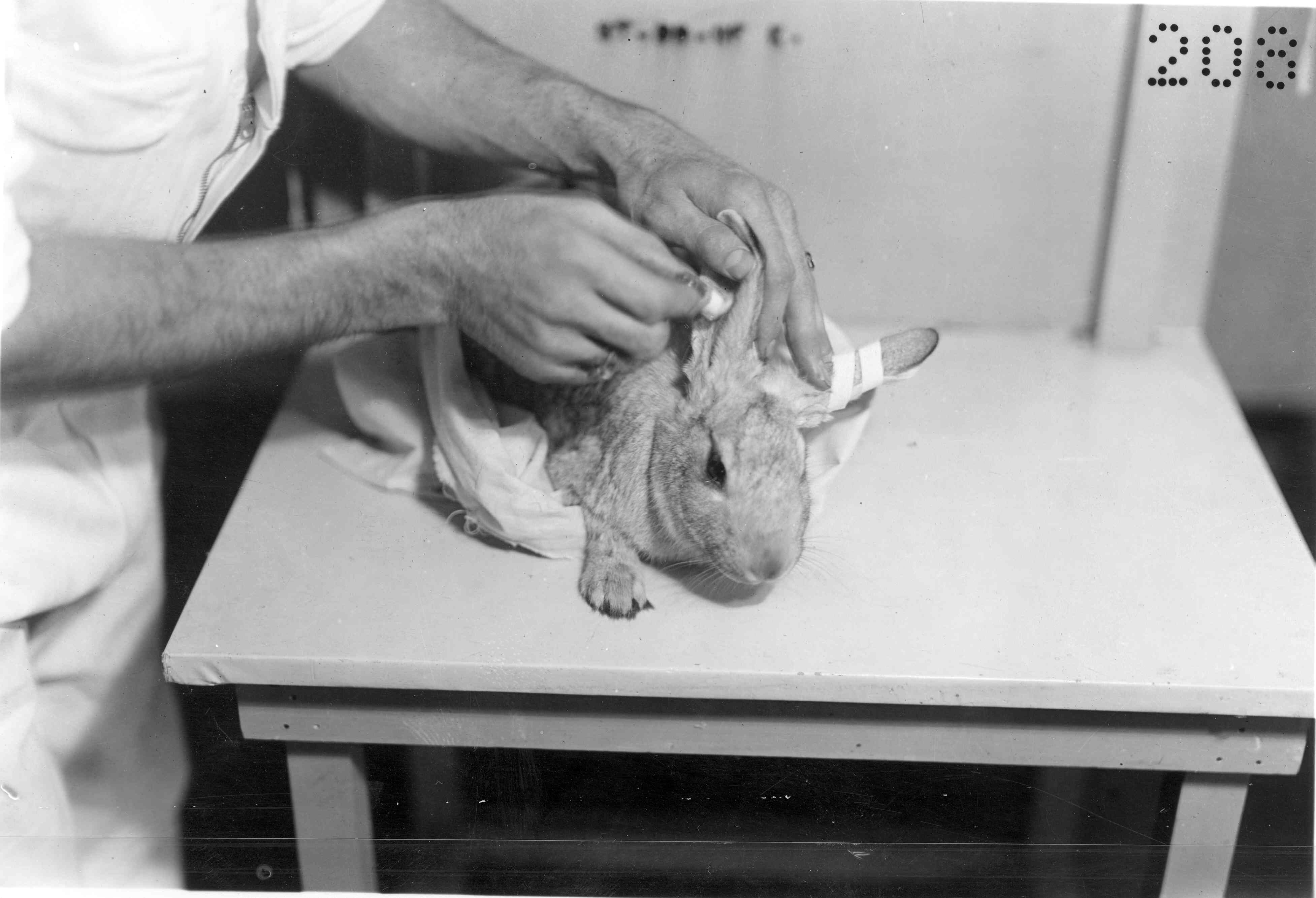
Image: Office of NIH History and Stetten Museum, 1478
2. Put the females put in separate pill boxes, give the box a lot number, and place the box over moist sand so they can lay eggs.
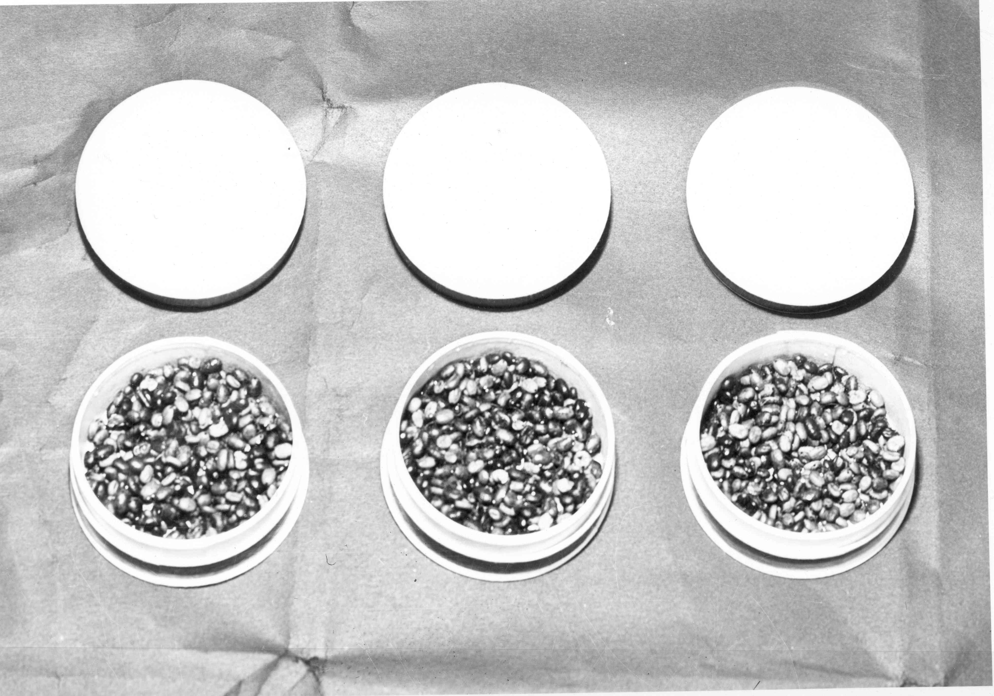
Image: Office of NIH History and Stetten Museum, 1487-1
3. After the eggs hatch, feed the whole group of larvae on infected rabbit. Do the same after they molt and become nymphs (as shown in the tubes) on a non-infected rabbit.
4. After each feeding, inject a few ticks into a guinea pig’s abdomen to see if the guinea pig gets RMSF. Then you know the ticks are infected too.
5. After the ticks molt to become adults, keep them a month to let the RMSF infection grow in them.
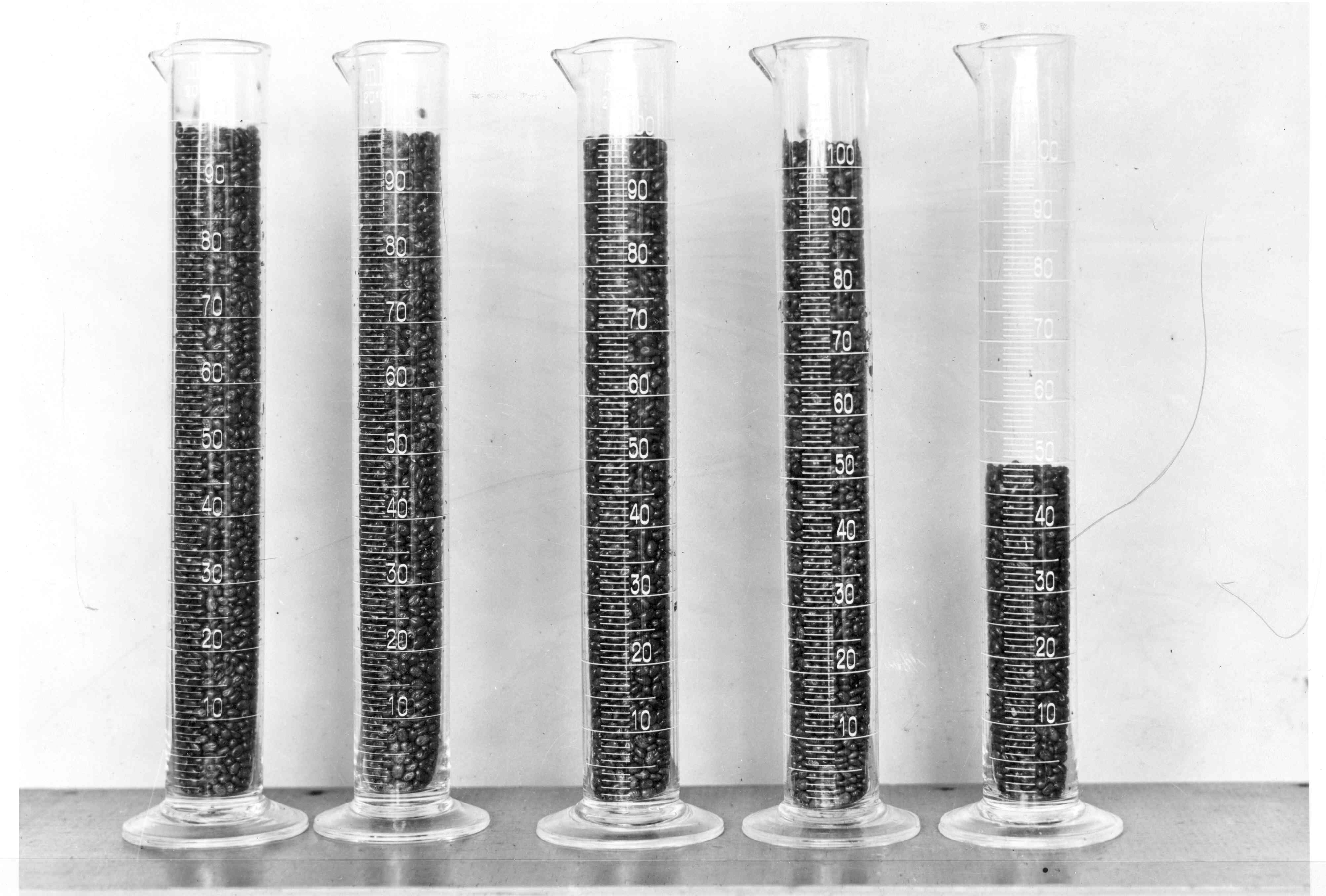
Image: Office of NIH History and Stetten Museum, 1486
6. Feed the adult ticks on an infected guinea pig. Then eviscerate the ticks and grind them in a mortar for 10-15 minutes with sterile sand and a bit of salt solution. This will separate their internal organs from their exoskeletons to create an emulsion.
7. Dilute the emulsion with salt solution so 1 cubic centimeter of emulsion equals two or more tick viscera.
8. Test the emulsion on two guinea pigs to find the minimal infectious dose. Both guinea pigs have to get RMSF to consider the emulsion for the vaccine.
9. Dilute the emulsion again to equal one tick. Add phenol so that the final product has 0.5% phenol (preservative-disinfectant).

Image: Montana Memory, 746
10. Let the mixture sit at room temperature for two or three days. A heavy precipitate is formed as seen in the photo, and extraneous organisms are killed as shown by anaerobic and aerobic sterility tests.
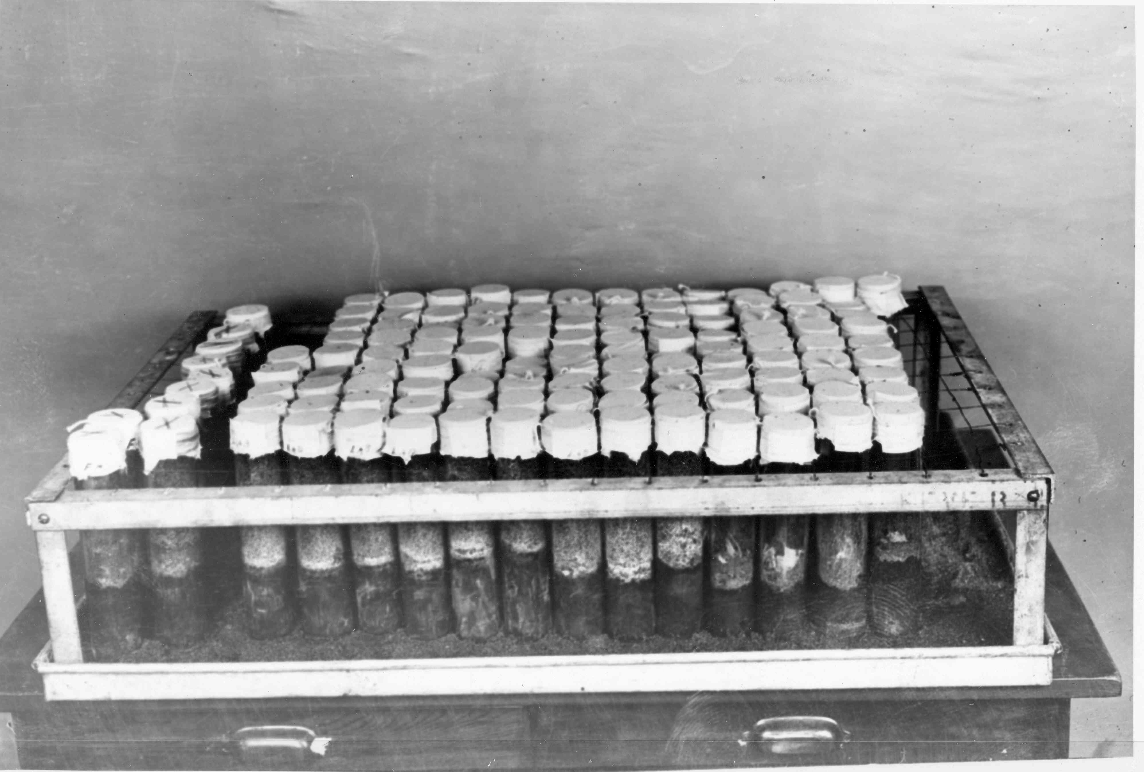
Image: Office of NIH History and Stetten Museum, 1477
11. Use a centrifuge to separate the precipitate, as Emily Emmart is doing in the photo, because it doesn’t go through filter paper, and the vaccine’s potency is destroyed by a Berkefeld filter. The supernatant fluid (clear fluid) is the vaccine.
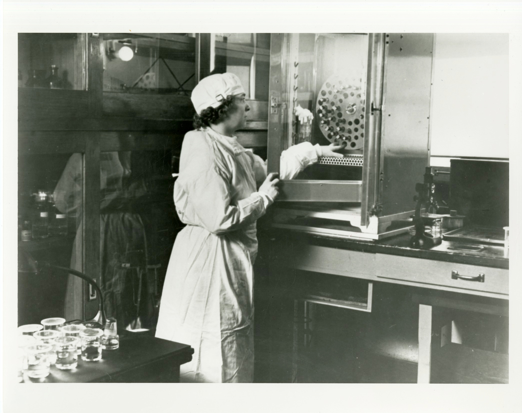
Image: National Library of Medicine, 101447516
12. Bottle the vaccine in a sterile environment.
The dosage is difficult to determine, but “Such irregularities are not surprising, however, when we recall that so little is known of the various factors affecting the process and mechanism of immunity.” (1925, page 2160 Monkeys).
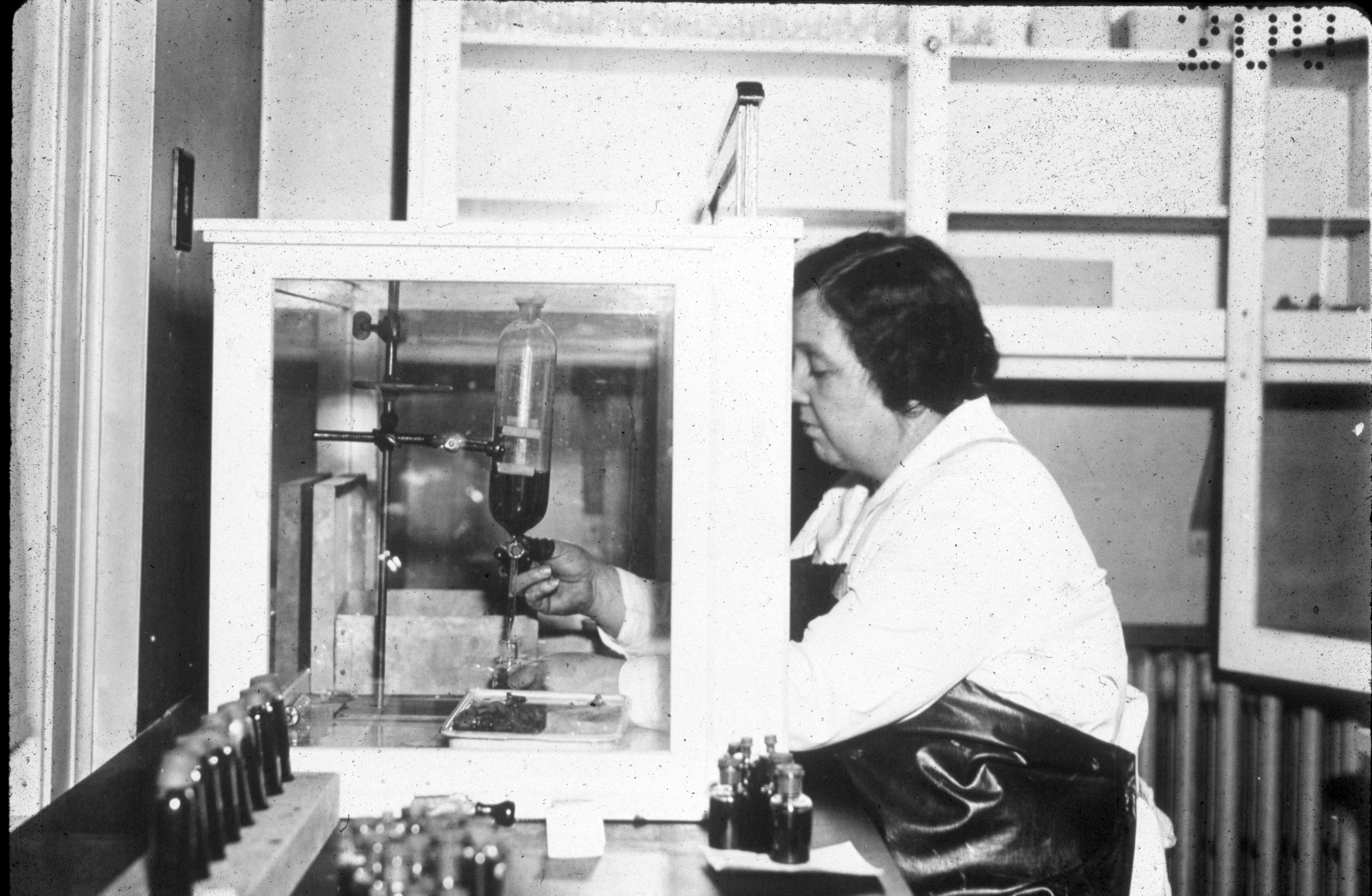
Image: Office of NIH History and Stetten Museum, 1523-sl
Giving Vaccinations
After using the Canyon Creek Schoolhouse laboratory staff as an informal clinical trial, vaccination trials of the people most exposed to RMSF began. People most likely to get RMSF because of their jobs were among those first vaccinated, such as shepherds and cattlemen. These shepherds in Idaho received their shots in 1926.
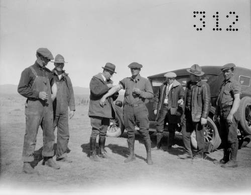
Image: Montana Memory, 312
The residents of the Bitterroot Valley, Montana, where the most people died from RMSF, were also vaccinated. In the photo, families lined up outside a local school in Darby to be vaccinated. Dr. R. R. Hayward, a local physician (in a white lab coat), administered the vaccine to a man in his left arm. The town of Darby had lost Arthur Kerlee to the disease when he was working at the Canyon Creek Schoolhouse laboratory.
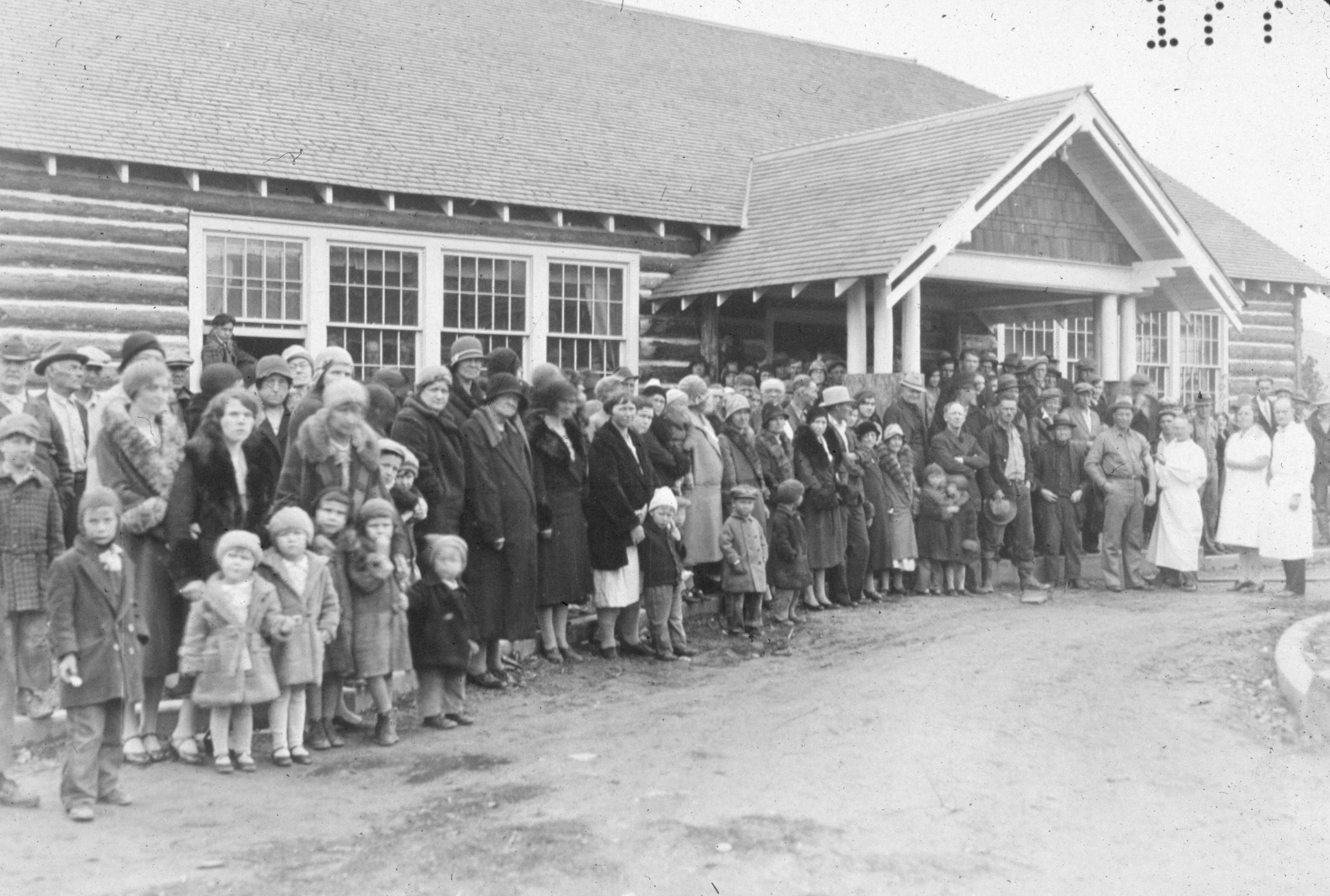
Image: Office of NIH History and Stetten Museum, 1524-sl
In this photo, vials of the Spencer-Parker RMSF vaccine sit on the letter or telegram requesting the vaccine from places all over the United States. The Canyon Creek Schoolhouse laboratory was producing as much of the vaccine as possible, but demand for the vaccine exceeded the supply, making the construction of a new building designed specifically for scientific research and vaccine production necessary—Building One of the Rocky Mountain Laboratories.
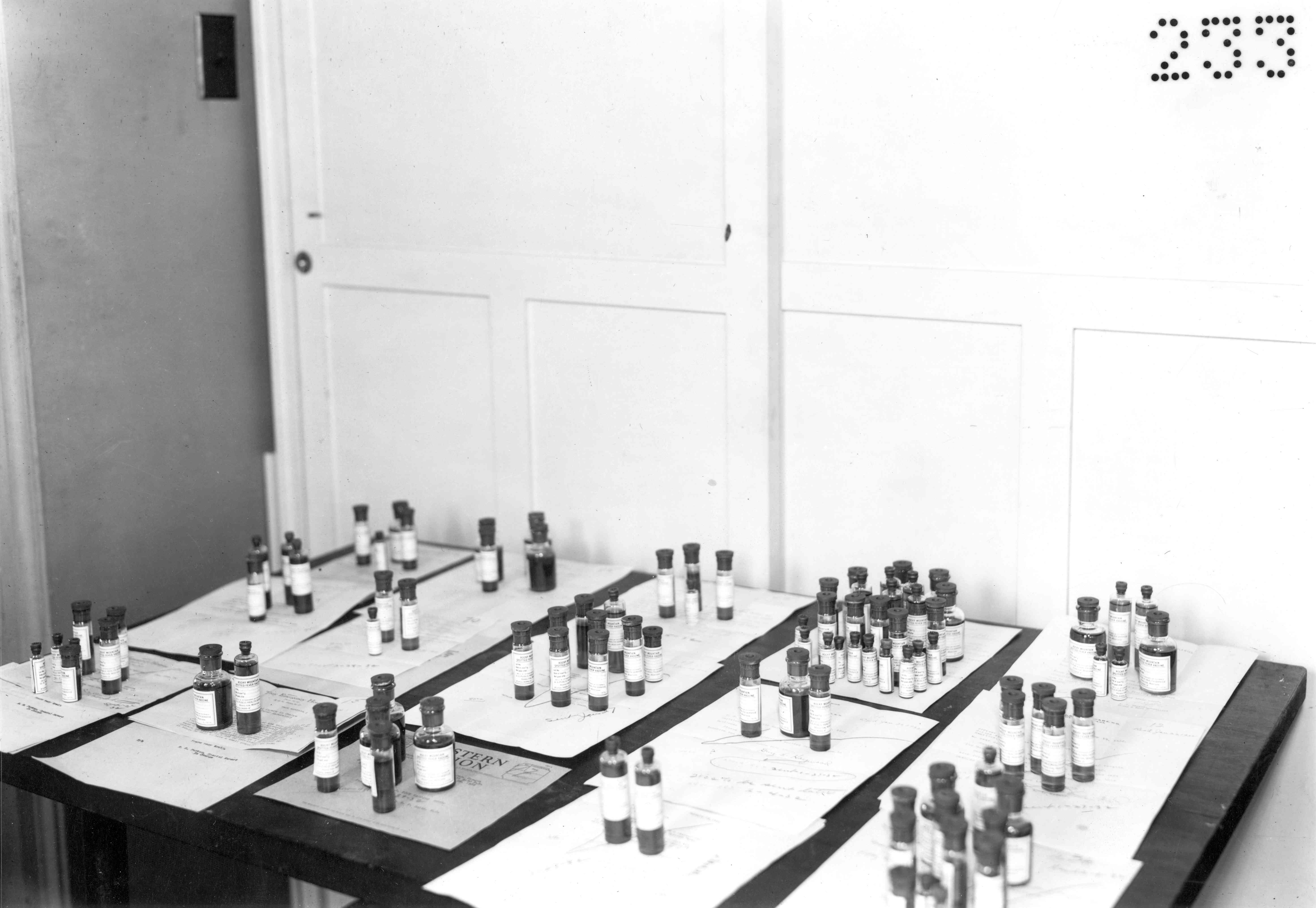
Image: Office of NIH History and Stetten Museum, 1560
Can We Treat or Cure RMSF?
A Vaccine Made Unnecessary
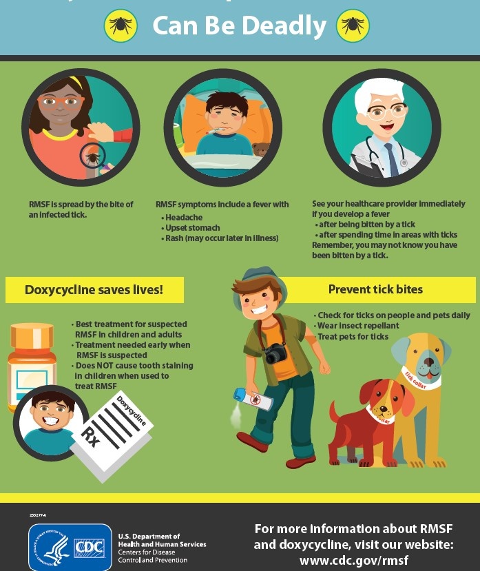
One question not answered at the Canyon Creek Schoolhouse laboratory was if there was an effective treatment for Rocky Mountain spotted fever. But in the late 1940s, antibiotics were found to cure the disease; this discovery made the Spencer-Parker vaccine obsolete. This current infographic from the CDC reminds us that RMSF is still a threat to people’s health. Visit their website for more information https://www.cdc.gov/rmsf/index.html
Overview
Content Tools
ThemeBuilder