
Santiago Ramón y Cajal Exhibit
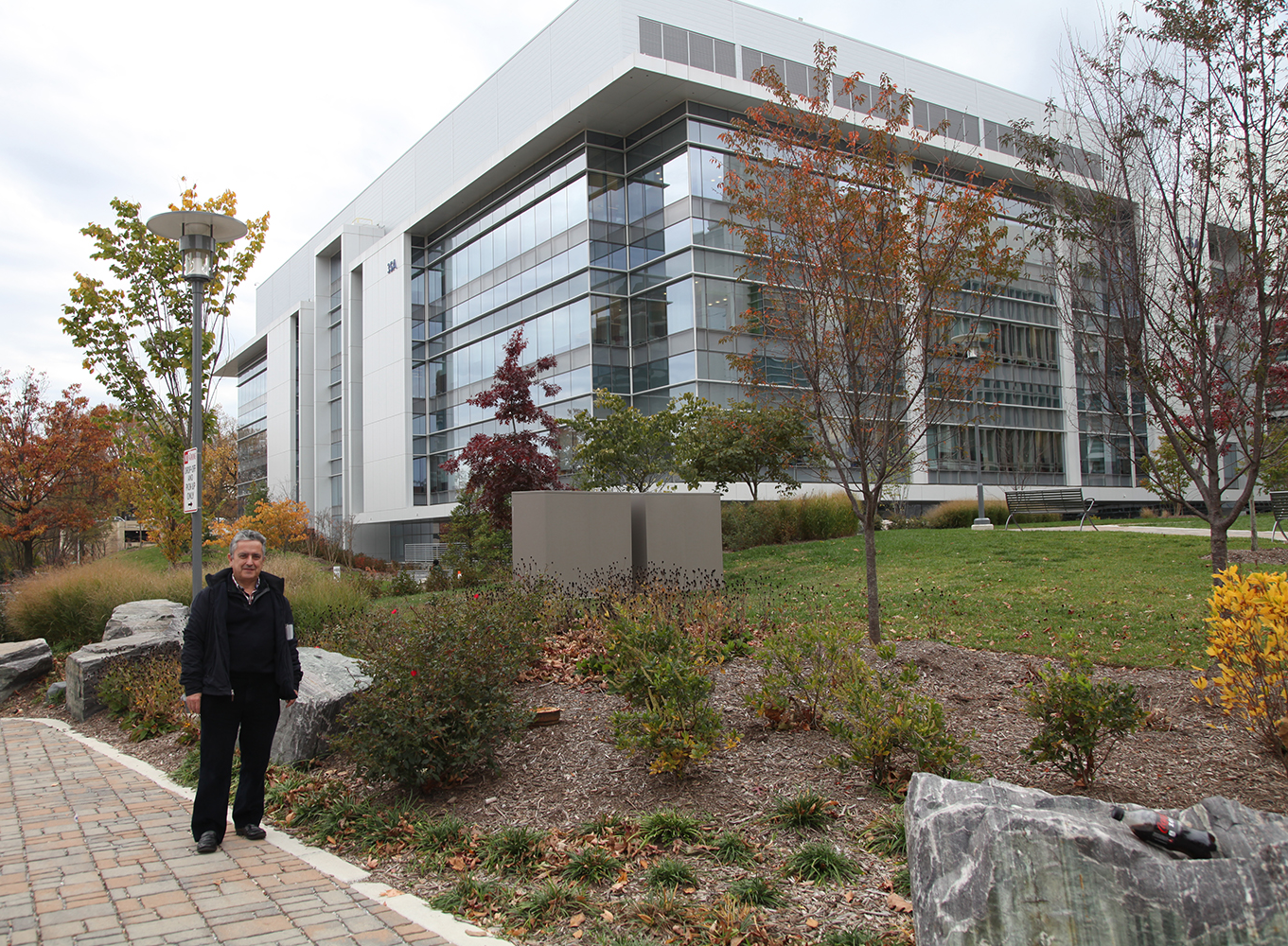
Ricardo Martínez Murillo, Ph.D., Director of the Instituto Cajal in Madrid, Spain, is pictured in front of NIH's recently dedicated neuroscience research center where the exhibition of Ramón y Cajal original drawings is located.
On March 31, 2014, the second phase of the John Edward Porter Neuroscience Research Center was dedicated. This new facility is shared by scientists from the National Institute of Neurological Disorders and Stroke (NINDS), National Eye Institute (NEI), National institute of Child Health and Human Development (NICHD), National Institute of Dental and Craniofacial Research (NIDCR), National institute of Mental Health (NIMH), National Institute on Deafness and Other Communicable Disorders (NIDCD), and the National Institute of Biomedical Imaging and Bioengineering (NBIB)—and represents a unique opportunity for scientists to collaborate across academic disciplines as well as the boundaries that sometimes separate institutes and centers on the NIH campus.
A new wave of research and exploration is beginning within these walls with new support for the creation of a new arsenal of instruments for unlocking the mysteries of the brain through the Brain Research through Advancing Innovative Neurotechnologies (BRAIN) Initiative. This moment may be the most appropriate to look back over the accomplishments of the last century and anticipate those of the next.
Santiago Ramón y Cajal
May 1, 1852 – October 17, 1934
Santiago Ramón y Cajal, a Spanish physician and scientist, was the first to describe the structure of the nervous system with exquisite precision. In what would become known as the “neuron doctrine,” he showed that the nervous system comprises individual cells (later termed “neurons”), that these cells connect to each other at small, specialized contact zones (now known as “synapses”), and that a single nerve cell typically possesses three anatomically distinct structures: the dendritic arbor, the cell body, and the axon. He further posited that neurons function as information processing units, using electrical impulses to communicate within functional networks. Cajal’s experimental work and theories provided the foundation for modern neurobiology.
An exhibition featuring revolving sets of seven original illustrations of famed scientist/artist Santiago Ramón y Cajal (on loan from the Instituto Cajal in Madrid, Spain), may be found near the North Entrance, on the first floor, of Building 35 on the NIH Campus.
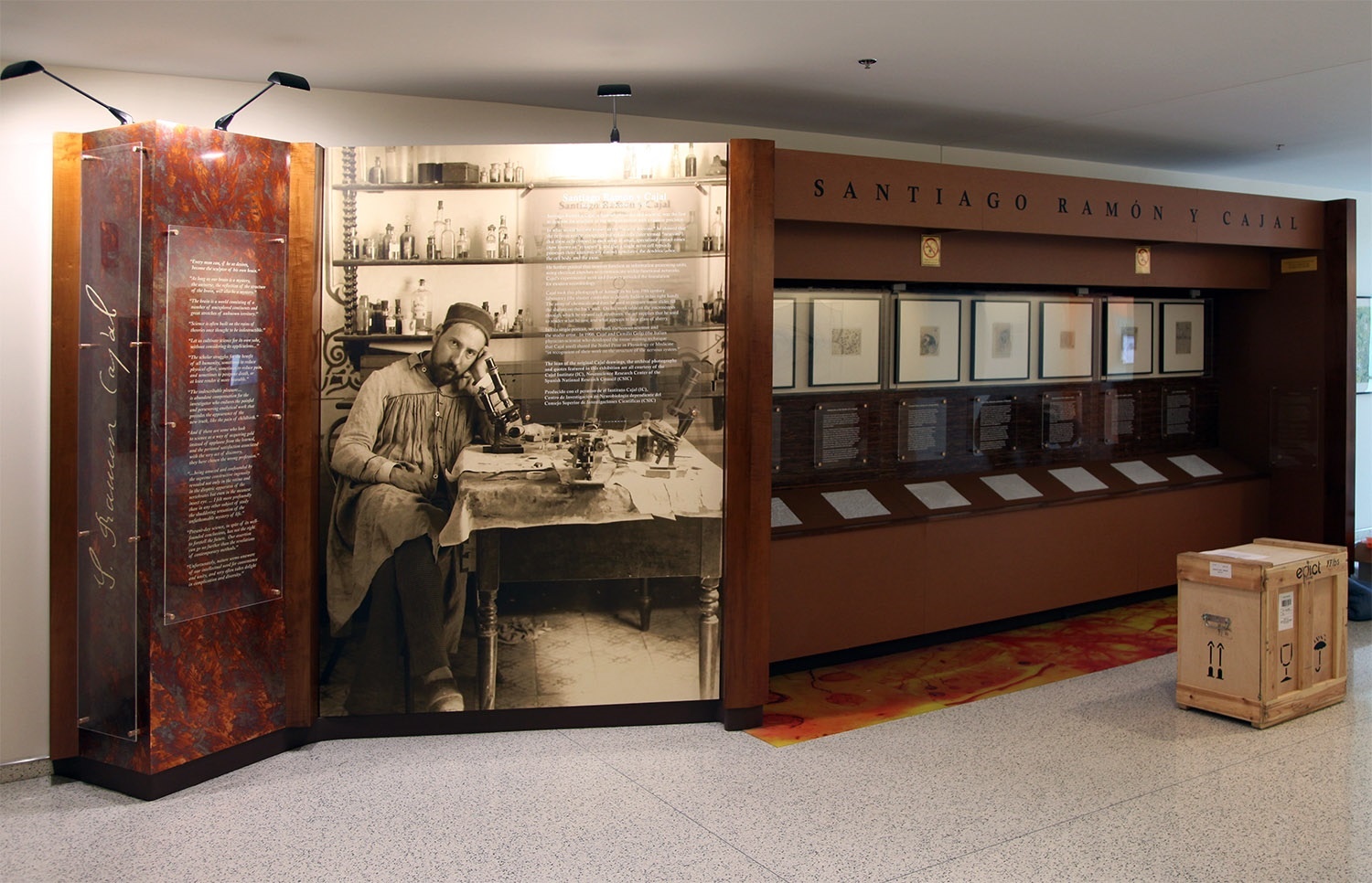
Cajal took this photograph of himself in his late-19thcentury laboratory (the shutter controller is cleverly hidden in his right hand). The array of chemicals and dyes he used to prepare tissue slides fill the shelves on the back wall. On his work table sit the microscopes through which he viewed cell structures, the art supplies that he used to render what he saw, and what appears to be a glass of sherry. In this single portrait, we see both the serious scientist and the studio artist. In 1906, Cajal and Camillo Golgi (the Italian physician-scientist who developed the tissue staining technique that Cajal used) shared the Nobel Prize in Physiology or Medicine “in recognition of their work on the structure of the nervous system.”

Each featured original illustration from the early 1900s, is accompanied by a caption written to engage scientists, researchers and investigators who populate the NIH campus. As well as a 3-D printed rendering that enlarges a detail of the illustration above. In this way, the drawings are rendered more accessible to a variety of audiences—including vision-impaired visitors who can directly experience these tactile versions of Cajal's drawings. These files are made available on the 3D Print Exchange. Direct links to the 3-D print files are provided at the end of this page.
The Cajal illustrations currently on-view include:
- Auditory Tracts
- Axonal Tracts in Rat
- Cajal-Astrocytes
- Cajal Astrocytes
- Cerebral Cortex-ii
- Interneuronal Plexuses
- Medulla
3D Printed Files
4th Installation (current)
- 3dpx-010423 - Auditory Tracts
- 3dpx-010424 - Axonal Tracts in Rat
- 3dpx-010425 - Cajal-Astrocytes
- 3dpx-010426 - Cajal-Astrocytes
- 3dpx-010427 - Cerebral cortex-ii
- 3dpx-010428 - Interneuronal Plexuses
- 3dpx-010429 - Medulla
3rd Installation
Unknown macro: {span}
Credit Line: Sorry folks, but I can't seem to solve the "adding a macro" step. The correct credit line for all of the original Cajal drawings is as follows:
"Cajal Institute, Spanish National Research Council or CSIC©"
2nd Installation
Calyx of Held
The Calyx of Held, first described by Hans Held in 1893, is one of the largest synapses found in the mammalian brain. As part of the auditory system, each Calyx is part of the axon of a globular bushy cell in the anteroventral cochlear nucleus, which forms a synapse with a principal cell in the medial nucleus of the trapezoid body. These synapses are integral to detection and localization of high frequency sounds.
At a time when the existence of synapses between nerve cells were not yet accepted as fact, Cajal’s drawings of the mammalian auditory system revealed a sophisticated understanding of the relationship between the Calyx of Held and the neuronal cell body it envelops. Rather than portraying[WC([1] them as a single, connected entity, Cajal’s coloring of this drawing indicates that he knew each part belonged to a separate cell, and that information would need to travel between them.
Because of the large size of a Calyx of Held and its relative accessibility, it has been a popular model system for neurobiological research. In particular, its amenability to patch-clamping has made it a favorite for systematic studies of presynaptic mechanisms, which can be involved in neurological disorders such as Parkinson’s disease.
- 3DPX-002127 - Developing Neocortex
- 3DPX-002126 - Olfactory System
- 3DPX-002125 - Insect Visual System
- 3DPX-002123 - Information Flow in Retina
- 3DPX-002116 - Astrocytes
1st Installation
3211
This drawing by Cajal appears to be a closer view of the dentate gyrus and the CA3 region of the Horn of Ammon shown in 3123. The narrower focus of this piece allowed Cajal to depict a wealth of detail in both the neurons themselves and the connections between them. The axons of the neurons in the dentate gyrus delicately entwine around the dendrites of the giant pyramid cells in the Horn of Ammon, while the axons of the giant pyramid cells stretch toward the fimbria, relaying information onwards. Both of Cajal’s major contributions to the field of neuroscience are on display here: the separate, yet intertwined cells make it clear that Neuron Theory prevails, while the arrows signify what Cajal intuited about the arrangement of the axons and dendrites and the direction of signal transduction, illustrating his law of ‘Dynamic Polarization’.
3123
Cajal inferred the flow of information between neurons from the shapes he saw on his slides. His ‘Law of Dynamic Polarization’ states that each neuron is polarized: it has dendrites, through which signals are received, and an axon through which signals are transmitted to the dendrites of the next cells. Thus, simply by observing the position of neurons in a tissue, he was able to discern the direction of signal transmission. While this was relatively easy in a tissue like the retina, where outside stimuli arrive from a fixed direction and must be carried inward, his real genius was shown in the deduction of the information flow in a complex tissue such as the hippocampus where output and input are not immediately obvious. The hippocampus, named for its resemblance to a genus of seahorses, consists of the Horns of Ammon (cornu ammonus), the dentate gyrus, and the subiculum. It receives signals from various parts of the brain via the enterorhinal cortex, which travel through the hippocampus in the path shown by Cajal above, and its output travels through the fornix to the anterior thalamic nuclei, among other destinations. Previously to Cajal’s observations, it was thought that the fornix was a source of input for the hippocampus. Due to the cellular organization of the hippocampus, where the dendrites of many pyramidal neurons run parallel to each other and contain spines that amplify input, it is often a focal point for epileptic seizures, in which large numbers of pyramidal cells fire synchronously, disrupting the normal electrical activity of the region.
11308
Cajal observed that the axons of motor neurons were always at the end of the neuron facing away from the brain. Although he did not know the nature of the signal being sent, Cajal hypothesized that the command to move began in the motor cortex of the brain and traveled down the spinal cord to the muscles involved. In the same way, he noticed that the axons of sensory neurons were always on the end closer to the brain, and thus inferred that sensory input travels up the spinal cord to be processed by the brain. These observations were instrumental in the formulation of his ‘Law of Dynamic Polarization’. This work formed the basis for the work of Sherrington, winner of the 1932 Nobel Prize in Medicine, who developed a theory about the dynamics of nervous transmission based on Cajal’s drawings with arrows showing the direction of signal transmission; Sherrington hypothesized that the reflex responses he studied were based on anatomical circuits: nerves and the neurons of which they were comprised. The idea that one could trace a complete pathway through the nervous system inspired the work of Eric Kandel, winner of the Nobel Prize in 2000, who researched how sensory inputs from different body parts are connected to different parts of the brain.
11312
Cajal’s most well-known contribution to the field of neuroscience is the idea of and the supporting evidence for neuron theory: the idea that the nervous system is composed of individual cells. Before his precise observations, scientists generally believed in the reticular theory: that the central nervous system was made up of a continuous network of cells, allowing fast transduction of nervous signals within the syncytium, and that any observed free nerve endings in the peripheral nervous system were purely for receiving outside stimuli. Using the silver-chromate staining technique pioneered by Golgi, with whom he later shared the Nobel Prize, Cajal was able to see that cells in the central nervous system of vertebrates had free endings and were often intertwined with many different neuronal cell types, suggesting that each nerve cell was a separate entity. Golgi himself favored the reticular theory, even after Cajal published evidence to the contrary; in the diagram above, Cajal makes a direct comparison between Golgi’s ideas and what he himself saw under the microscope — instead of a network of connected cells, there were separate cells in close proximity. While neuron theory became widely accepted after Cajal’s findings were published in the late 1880s, Golgi and others continued to espouse the reticular theory into the early 1900s. It was not until 1954 that electron microscopy by Palade and Palay definitively demonstrated the existence of synapses and thus, the separation between neurons.
11322
By the time Cajal began staining his sections, scientists had been studying the gross structure of the cerebellum for almost 300 years and various sub-structures had been recognized, such as the lingula and the deep nuclei. Cajal’s elegant drawings identified a variety of cell types present in the cerebellum; in particular, the mossy and climbing fibers were unknown before, and their structure and placement suggested intriguing connections between the cerebellum and other parts of the brain. The technology to test any such connections would not be available for several decades, but in the 1960s, John Eccles was able to map the circuitry of the many types of cerebellar neurons using electrophysiological analysis. He found that the cerebellum differs from other parts of the brain in that nearly all signal transduction is unidirectional: the mossy fibers receive sensory signals from different parts of the spinal cord, which are transmitted through the granule cells to the Purkinje cells, while climbing fibers transmit signals from the inferior olivary nucleus to the Purkinje cells. The Purkinje cells in turn transmit the signals on to the deep nuclei, outside the cerebellar cortex, which eventually relay information to the cerebral cortex. Studies of subjects with damaged cerebellums suggested that the main function of the cerebellum was in fine-tuning movements; more recent research has revealed that the cerebellum also plays a role in the control of some non-motor functions, such as spatial cognition, working memory, and language use.
3818
Cajal’s careful studies of retinal sections from a variety of animals led him to conclude that retinal structure is very similar between species: all vertebrates have rod and cone photorecptors, as well as bipolar cells, amacrine cells, horizontal cells, interplexiform cells, and ganglion cells, in roughly the same arrangement. The vertebrate retina develops as an outgrowth of the brain, and signals received from stimulation by light travel through the retinal network through one of 3 now-established pathways and then into the brain through the optic nerve. The clearly implied signal transduction pathway of the light signal through the retina, from the photoreceptors through the ganglion cells, was one of Cajal’s initial inspirations for his ‘dynamic polarization’ theory. Further studies of the retina led to the Nobel Prize in Medicine in 1967 for Granit, Hartline, and Wald, for their respective discoveries of the electrophysiological properties of retinal neurons, lateral inhibition among neurons, and the mechanism of rhodopsin function. Later still, William Hagins at the NIH discovered that vertebrate retinal cells are depolarized in the dark, and become hyperpolarized when exposed to even a single photon, demonstrating the robustness of the signalling pathway hypothesized by Cajal so long ago.
11328
Neuron theory was advanced through tireless promotion of Cajal’s stained neural sections, in which distinct cell boundaries were clearly visible. Cajal was able to detect the delicate structure of neurons, which had previously been impossible, by combining Golgi’s silver nitrate staining with the ‘ontological method’: the use of samples from embryological or perinatal tissue, in which the neurons were unmyelinated and thus more easily susceptible to staining. This section of the cerebral cortex from a human infant is an excellent example of the success of his technique. Though their processes overlap, the cells are clearly distinct from one another. By noting the placement of the axons and dendrites, Cajal was also able to postulate the direction in which information flows through this tissue --- from the deeper layers of the cortex up toward the surface. The precentral gyrus is now known to be part of the primary motor cortex, which coordinates with several other parts of the brain to plan and execute muscle movements.
Test Print
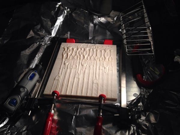
Photograph of the 3D printed plates being glued together
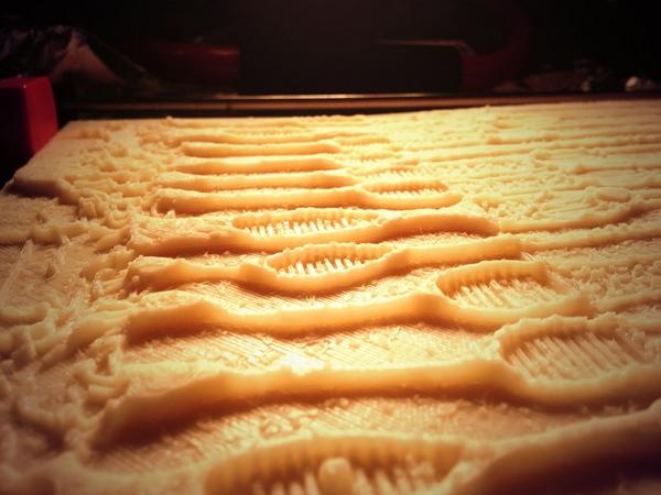 Close up of the 3D print
Overview
Content Tools
ThemeBuilder