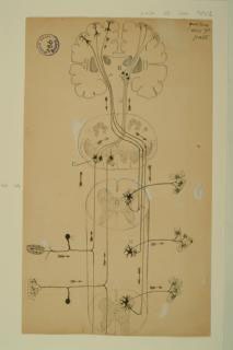
Cajal observed that motor neuron axons originated at the end of the neuron facing away from the brain. Although he did not know the nature of the signal being sent, Cajal hypothesized that the command to move began in the motor cortex of the brain and traveled down the spinal cord to the muscles involved.
In the same way, he noticed that the axons of sensory neurons originated at the end of the neuron that was closer to the brain and inferred that sensory input travels up the spinal cord to be processed by the brain.
These observations were instrumental in the formulation of his ‘Law of Dynamic Polarization,’ which was extended further by Sir Charles Sherrington, winner of the 1932 Nobel Prize in Physiology or Medicine, who showed that spinal cord reflex circuits involved “reciprocal innervation” of opposing muscles.
Cajal’s greatest contribution to neuroscience is the idea of and supporting evidence for the “neuron doctrine:” the nervous system is composed of individual cells.
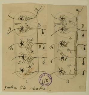
Before his precise observations, scientists generally held the reticular theory, championed by Camillo Golgi, who argued that the central nervous system was a continuous network of cells, allowing fast transmission of nerve signals across a large syncytium, and that any observed free nerve endings in the peripheral nervous system were purely receivers of outside stimuli.
Using the silver chromate staining technique pioneered by Golgi, with whom he later shared the Nobel Prize, Cajal observed that cells in the vertebrate central nervous system had free endings that were often apposed to many different neuronal cell types, suggesting that each nerve cell was a separate entity.
In the diagram shown here, Cajal compared Golgi’s theory of continuity (I, left) with the contiguity that he observed under themicroscope (II, right). Cajal’s neuron doctrine was widely accepted after he published his findings in the late 1880s. It was not until the 1950s that electron microscopy by Palade and Palay definitively demonstrated the existence of synapses and the physical separation between neurons.
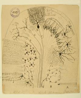
Scientists had studied the gross structure of the cerebellum for almost 300 years, recognizing various sub-structures, before Cajal identified a variety of cell types present in the cerebellum; the mossy and climbing fibers were unknown before, and their structure and placement suggested intriguing connections between the cerebellum and other parts of the brain.
In the 1960s, Sir John Eccles used electro-physiological analyses to reveal elegant feedforward circuitry. Incoming mossy fibers deliver sensory signals from pre-cerebellar nuclei and the spinal cord to granule cells (g), the most numerous neuron type in the brain. Granule cell axons bifurcate to form the densely arrayed parallel fibers, which contact the (relatively) giant Purkinje cells (a).
Meanwhile, climbing fibers transmit signals from the inferior olivary nucleus to Purkinje cells. Each parallel fiber makes a single weak synapse onto a Purkinje cell, whereas the parallel fiber connection is very strong, comprising hundreds of distinct synaptic contacts.
Purkinje cells transmit inhibitory signals to the deep nuclei, outside the cerebellar cortex, which will relay information to the cerebral cortex. The cerebellum coordinates and fine-tunes movements, although recent research has revealed roles for the cerebellum in spatial cognition and language.
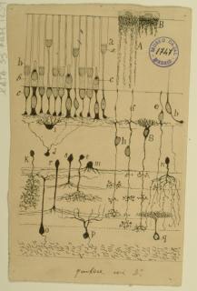
Cajal’s careful studies of retinal sections from a variety of animals led him to conclude that retinal structure is remarkably similar between species: all vertebrates have rod and cone photoreceptors, as well as bipolar cells, horizontal cells, amacrine cells and ganglion cells, in roughly the same arrangement.
The vertebrate retina develops as an outgrowth of the brain, and signals received from stimulation by light travel through the retinal network and then into the brain via the ganglion cell axons in the optic nerve. The clear signaling pathway through the retina, from the photoreceptors through the ganglion cells, was one of Cajal’s initial inspirations for his ‘dynamic polarization’ theory.
Further studies of the retina led to the Nobel Prize in Medicine in 1967 for Granit, Hartline, and Wald, for their respective discoveries of the electrophysiological properties of retinal neurons, lateral inhibition among neurons, and the mechanism of rhodopsin function.
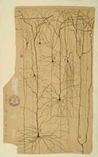
Neuron theory was advanced through tireless promotion of Cajal’s stained brain sections, in which distinct neuron boundaries were clearly visible. Cajal was able to detect the delicate structure of neurons by applying Golgi’s silver nitrate staining method to samples from embryological or perinatal tissue, in which the neurons were unmyelinated and thus more easily susceptible to staining.
This section of the cerebral cortex from a human infant is an excellent example of the success of his technique. Though their processes overlap, the cells are clearly distinct from one another. By noting the placement of the axons and dendrites, Cajal was also able to postulate the direction in which information flows through this tissue—from the deeper layers of the cortex up toward the surface.
The precentral gyrus is now known to be part of the primary motor cortex, which coordinates with several other parts of the brain to plan and execute muscle movements.