Versions Compared

Key

  • This line was added.
  • This line was removed.
  • Formatting was changed.

...

Div
classgrid-row grid-gap
Div
classdesktop:grid-col-6:desktop

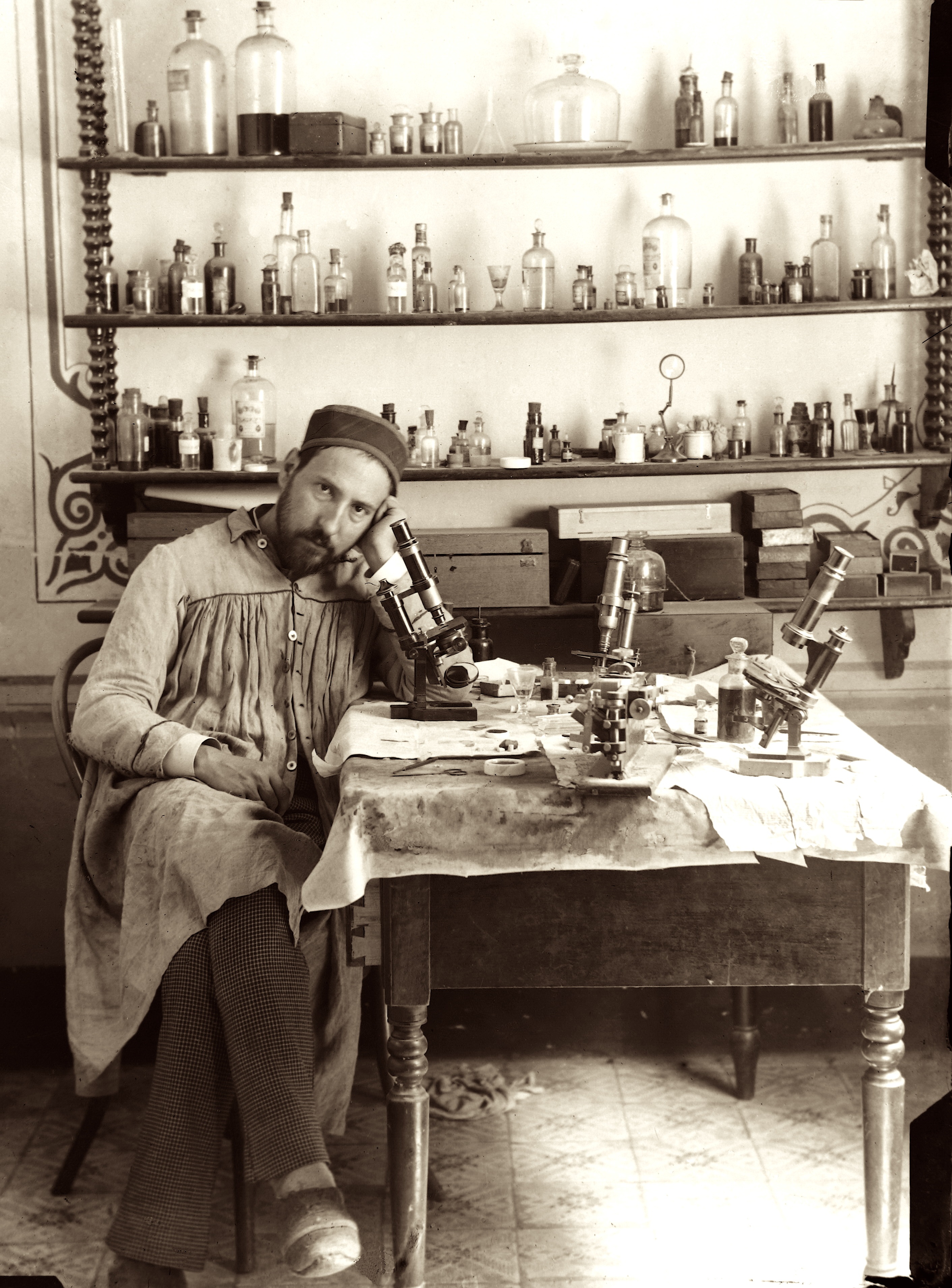
Photograph of Santiago Ramón y CajalSantiago Ramón y Cajal

Span
classcredit

Courtesy of the Cajal Institute, Spanish National Research Council or CSIC©

Div
classdesktop:grid-col-6:desktop

Santiago Ramón y Cajal, a Spanish physician and scientist, was the first to describe the structure of the nervous system with exquisite precision.  In what would become known as the “neuron doctrine,” he showed that the nervous system comprises individual cells (later termed “neurons”), that these cells connect to each other at small, specialized contact zones (now known as “synapses”), and that a single nerve cell typically possesses three anatomically distinct structures: the dendritic arbor, the cell body, and the axon. He further posited that neurons function as information processing units, using electrical impulses to communicate within functional networks.  Cajal’s experimental work and theories provided the foundation for modern neurobiology.

An exhibition featuring revolving sets of seven original illustrations of famed scientist/artist Santiago Ramón y Cajal (on loan from the Instituto Cajal in Madrid, Spain), may be found near the North Entrance, on the first floor, of Building 35 on the NIH Campus.

Cajal took this photograph of himself in his late-19thcentury laboratory (the shutter controller is cleverly hidden in his right hand). The array of chemicals and dyes he used to prepare tissue slides fill the shelves on the back wall. On his work table sit the microscopes through which he viewed cell structures, the art supplies that he used to render what he saw, and what appears to be a glass of sherry. In this single portrait, we see both the serious scientist and the studio artist. In 1906, Cajal and Camillo Golgi (the Italian physician-scientist who developed the tissue staining technique that Cajal used) shared the Nobel Prize in Physiology or Medicine “in recognition of their work on the structure of the nervous system.” 

...

Div
classgrid-row grid-gap
Div
classdesktop:grid-col-6:desktop

Each featured original illustration from the early 1900s, is accompanied by a caption written to engage scientists, researchers and investigators who populate the NIH campus.  As well as a 3-D printed rendering that enlarges a detail of the illustration above.  In this way, the drawings are rendered more accessible to a variety of audiences—including vision-impaired visitors who can directly experience these tactile versions of Cajal's drawings.  These files are made available on the 3D Print Exchange.  Direct links to the 3-D print files are provided at the end of this page.

The Cajal illustrations currently on-view include:

  • Auditory Tracts
  • Axonal Tracts in Rat
  • Cajal-Astrocytes
  • Cajal Astrocytes
  • Cerebral Cortex-ii
  • Interneuronal Plexuses
  • Medulla


Div
classdesktop:grid-col-6:desktop

Close up of Santiago Ramón y Cajal Exhibit illustration and 3d print

Div
classhr-3


Video

Widget Connector
width100%
urlhttps://www.youtube.com/watch?v=QIDUKHXqtg4
height560

Div
classhr-3


5th Installation (current)

Upper cervical ganglia

Div
classgrid-row grid-gap
Div
classdesktop:grid-col-6
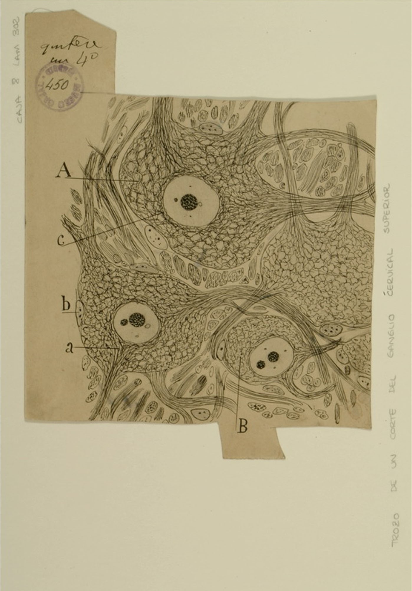
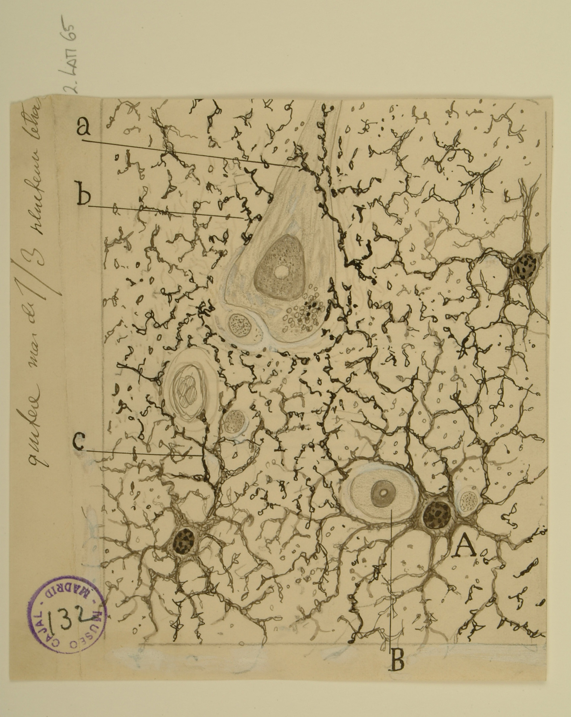
The autonomic nervous system controls involuntary body functions such as heart rate, digestion, and respiration, and is divided into two parts: the sympathetic nervous system and the parasympathetic nervous system. The sympathetic nervous system’s primary function is to activate the fight-or-flight response to danger, but it is also active at a basal level to preserve homeostasis. The superior cervical ganglion, a section of which is shown here, is a part of the sympathetic nervous system in which neurons originating in the spinal cord form synapses with neurons that innervate the heart, head, and neck, and control responses such as heart rate and pupil dilation. The cell bodies shown above are surrounded by “receptive nets” (A, B) formed by their own dendrites and the axons of neurons originating in the thoracic spinal cord. While Cajal himself performed preliminary work on the sympathetic nervous system, his former student, Fernando de Castro, gained international recognition for his careful studies of the fine structure of the autonomic ganglia. Cajal recognized de Castro as an equal and entrusted him with supervising the technical training and research of fellows at the Cajal Institute between 1924-1932.


Span
classusa-button

3dpx-010423 - Auditory Tracts

Div
classdesktop:grid-col-6

Span
classcredit

Courtesy of the Cajal Institute, Spanish National Research Council or CSIC©

...

Div
classgrid-row grid-gap
Div
classdesktop:grid-col-6

The superior temporal gyrus contains the auditory cortex, responsible for processing sound, and Wernicke’s area, which is necessary for the processing of speech to be understood as language rather than simply sounds. Shown here are several layers of pyramidal cells in the superior temporal gyrus, which is layered similarly to other areas of the temporal cortex. Though they vary in size and position, the pyramidal cells (a,b,c,d,e,f,g,h) all exhibit the characteristic cone-shaped cell body, a single apical dendrite extending upwards to the cortical surface, basal dendrites, and basal axons (a). Cajal characterized pyramidal cells from many tissues, detailing the variety of shapes and sizes found in different locations throughout the brain. He also hypothesized that size and shape of the dendritic arborizations of the pyramidal cells would vary over the lifespan of an organism depending; recent research reveals that the cells shown here, from the superior temporal gyrus of an infant, have much larger and denser dendritic branches than those from an adult would in this specific location.


Span
classusa-button

3dpx-010423 - Auditory Tracts

Div
classdesktop:grid-col-6

Span
classcredit

Courtesy of the Cajal Institute, Spanish National Research Council or CSIC©


Div
classhr-3


4th Installation

Nuclei in the auditory pathway

...

Div
classgrid-row grid-gap
Div
classdesktop:grid-col-6

The dorsal horn of the spinal cord contains an area known as both Rexed Lamina II and the substantia gelatinosa of Rolando, named for its gelatinous appearance due to a high concentration of small neurons and a lack of myelination.   Cajal classified the cells of the gelatinosa as either “limiting” (b, c) or “central” (d) according to their location within the area, their size, and their dendritic organization; modern neuroanatomists now classify the cells as “stalked” (b,c) or “islet”(d), although there are a variety of neurons in the gelatinosa that defy classification.  

Because the axons of many neurons in the gelatinosa appeared not to extend outside of the gelatinosa itself, it was once hypothesized that the gelatinosa was a “closed system;” we now know that neurons of the gelatinosa receive input from the spinothalamic tract and dorsal columns, and relay that information deeper into the spinal cord through Rexed Laminae III and IV.  

Neurons of the gelatinosa are unusually dense in Substance P and opioid-type pain receptors, and thus the gelatinosa is believed to play a role in the modulation/mediation of pain perception.


Span
classusa-button

3dpx-010429 - Medulla

Div
classdesktop:grid-col-6

Span
classcredit

Courtesy of the Cajal Institute, Spanish National Research Council or CSIC©


Div
classhr-3


3rd Installation

Cingulate Cortex 

Div
classgrid-row grid-gap
Div
classdesktop:grid-col-6
(vertical transverse section)

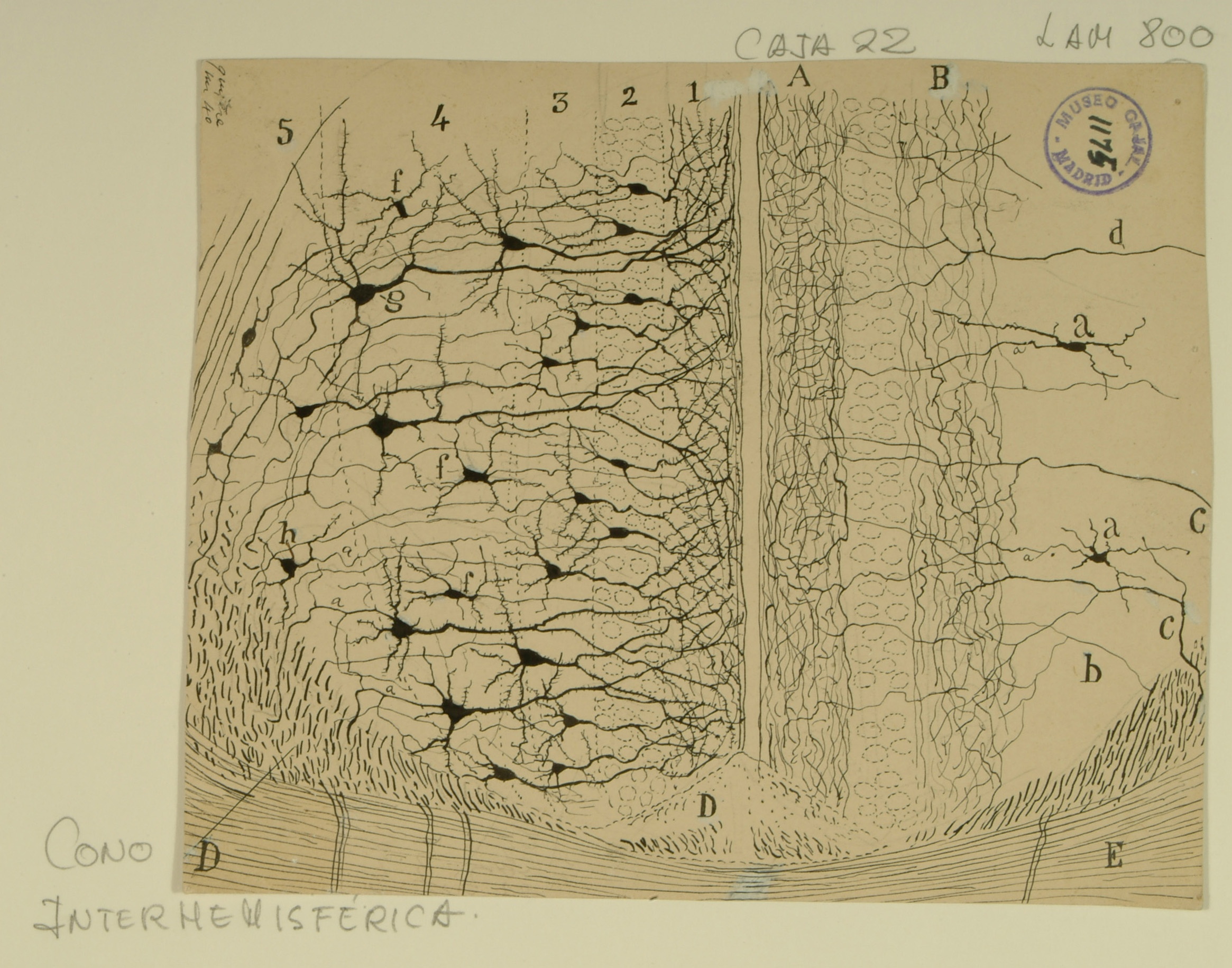
Among his many “firsts,” Ramon y Cajal was the first to analyze the structure of individual neurons in the cingulate cortex, a structure in the brain involved with emotion formation and processing, learning, and memory.  In this drawing, Cajal highlights the structure of the anterior cingulate cortex, as well as its surrounding tissues, the induseum griseum (right D), the cingulum bundle (left D), and the corpus callosum (E).  

While the layers of structure are intended to be the same on each side of the drawing, on the left side, Cajal emphasizes the pyramidal neurons present in the five cortical layers of the anterior cingulate cortex (1-5), while on the right side, he draws attention to the interneurons found in those layers (a, d).  In addition to the variety of types of pyramidal neurons that comprise the cingulate cortex (extroverted, small, medium, large, fusiform), there are also a variety of non-pyramidal neurons (multipolar, bipolar, and bitufted).  

The soma of the neurons and interneurons are mainly in layers 2-5, while layer 1 is made of a dense arbor of dendrites from the layers below.  Changes in the number, density, or composition of neurons in the anterior cingulate cortex are associated with disease states such as Parkinson’s disease, Alzheimer’s disease, and schizophrenia.


Span
classusa-button

3dpx-010430 - 3334

Div
classdesktop:grid-col-6

Span
classcredit

Courtesy of the Cajal Institute, Spanish National Research Council or CSIC©


...

Div
classgrid-row grid-gap
Div
classdesktop:grid-col-6

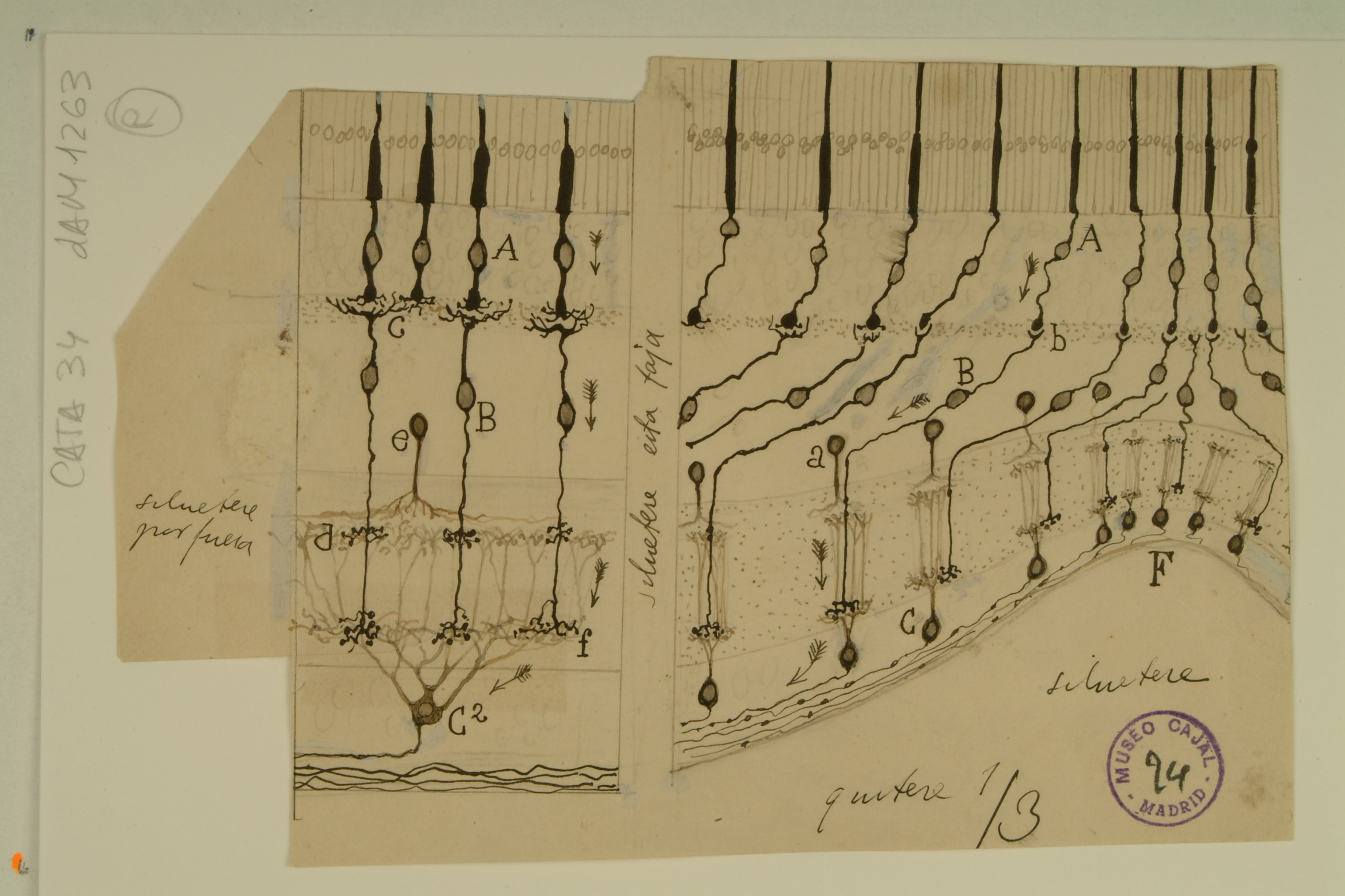
The fovea centralis, a small avascular depression at the center of the inner retinal surface filled with densely packed cone cells, is responsible for collecting visual information from the focus of visual gaze to form high-resolution images.  Signals from the fovea constitute half of all input to the visual cortex.  

In this drawing of the foveal pit (F) and perifoveal area by Cajal, the midget bipolar and ganglion cells (so called due to their small size) are highlighted.  In the image, each cone cell (A) contacts a single midget bipolar cell (B), which in turn makes synapses with a single midget ganglion cell (C), which then transmits information through the optic nerve to the visual cortex.  Cajal correctly noted that as distance from the foveal pit increases, the number of cone cells providing input to a single ganglion cell (C2) increases, a process known as convergence.  The lack of convergence at the foveal pit is what allows for maximum visual acuity.  

What Cajal did not realize was that, in order to maintain such visual acuity, each cone cell in the fovea is actually connected to two midget bipolar cells and midget ganglion cells (each pair of which exists in mutually exclusive planes of focus in microscope preparations) — one pair will signal only when the center of the cone’s photoreceptive area is stimulated, and the other pair signals only when the off-center area of the cone’s photoreceptive area is stimulated.  In addition, information transmitted from the fovea to the visual cortex also contributes to color vision.  


Span
classusa-button

3dpx-010436 - 11317

Div
classdesktop:grid-col-6

Span
classcredit

Courtesy of the Cajal Institute, Spanish National Research Council or CSIC©


Div
classhr-3


2nd Installation

Growth Cones

Div
classgrid-row grid-gap
Div
classdesktop:grid-col-6

The complicated circuitry of the brain and the precise connections of groups of neurons at various places within it suggest that neurons do not randomly form synapses as they grow, but instead respond to organizing signals.  Cajal was perhaps the first scientist to observe conical, fluid projections at the ends of developing neurons, and certainly the first to posit that such a structure might be involved in guiding the neuron towards a particular target.   

What we now know as growth cones – dynamic extensions of a developing neuron – respond to a variety of chemicals, secreted by target neurons and the extracellular matrix, that can be attractive or repulsive to the growth cone depending on the receptors present.  

The variety of the growth cones shown above is due to the complexity of the paths they were to navigate: the ones depicted in C were travelling a quick path through white matter in the brain, while those in A and B travelled a slower, more complicated path through gray matter and the ventral commissure, respectively.  

Current research has found that the shape of the growth cone in vivo differs somewhat from Cajal’s preserved specimens, but the complexity of the growth cone in relation to its travel speed remains the same.


Span
classusa-button

3DPX-002129 - Growth Cones

Div
classdesktop:grid-col-6

Span
classcredit

Courtesy of the Cajal Institute, Spanish National Research Council or CSIC©


...

Div
classgrid-row grid-gap
Div
classdesktop:grid-col-6

Once thought to be mere “filler” for the space around neurons, astrocytes are star-shaped glial cells found throughout the brain and spinal cord that are now known to perform many important functions, such as regulating the transmission of ions and glucose between blood vessels and the brain.  

When Cajal drew these protoplasmic astrocytes, the prevailing theory was that astrocytes only provided structural support for neurons.  He rejected this idea and instead hypothesized that all the astroglia in the brain formed a sort of gland, which would release substances that affect brain function; it is now known that astroglia do indeed release substances that affect neuronal signaling, including glutamate, GABA, and ATP.  

Having observed the close association of astrocytes with blood vessels and neurons, as well as the fact that all astrocytes appeared to have a prominent appendage, or “foot,” Cajal further hypothesized that astrocytes used this “foot” to stimulate blood vessel dilation.  Astrocyte “feet” do indeed regulate blood vessel diameter, albeit via the release of signaling molecules rather than through physical manipulation, as Cajal imagined.  Astrocyte-evoked changes in blood flow, and thus in oxygenation, constitute the signal that is measured by fMRI, a tool used to image brain activity.


Span
classusa-button

3DPX-002116 - Astrocytes

Div
classdesktop:grid-col-6

Span
classcredit

Courtesy of the Cajal Institute, Spanish National Research Council or CSIC©


Div
classhr-3


1st Installation

Dentate Gyrus and CA3 Region of the Hippocampus

...

Div
classgrid-row grid-gap
Div
classdesktop:grid-col-6

Neuron theory was advanced through tireless promotion of Cajal’s stained brain sections, in which distinct neuron boundaries were clearly visible.  Cajal was able to detect the delicate structure of neurons by applying Golgi’s silver nitrate staining method to samples from embryological or perinatal tissue, in which the neurons were unmyelinated and thus more easily susceptible to staining.  

This section of the cerebral cortex from a human infant is an excellent example of the success of his technique.  Though their processes overlap, the cells are clearly distinct from one another.  By noting the placement of the axons and dendrites, Cajal was also able to postulate the direction in which information flows through this tissue—from the deeper layers of the cortex up toward the surface.  

The precentral gyrus is now known to be part of the primary motor cortex, which coordinates with several other parts of the brain to plan and execute muscle movements.


Span
classusa-button

3DPX-000789 - Cortical Pyramidal Cells

Div
classdesktop:grid-col-6

Span
classcredit

Courtesy of the Cajal Institute, Spanish National Research Council or CSIC©


Div
classhr-3


Test Print

This initial print file was created to create a process for making a 3d printable file from the image data. Two things became apparent in the creation of the file.  One is that the relief, the distance between the background (generally paper without ink) and the foreground, neurons and structures inked on the paper, needed to be minimal in order to more effectively communicate the subject through touching with fingertips. If the relief was too high, as in this test print, then it became harder to feel small details.

...