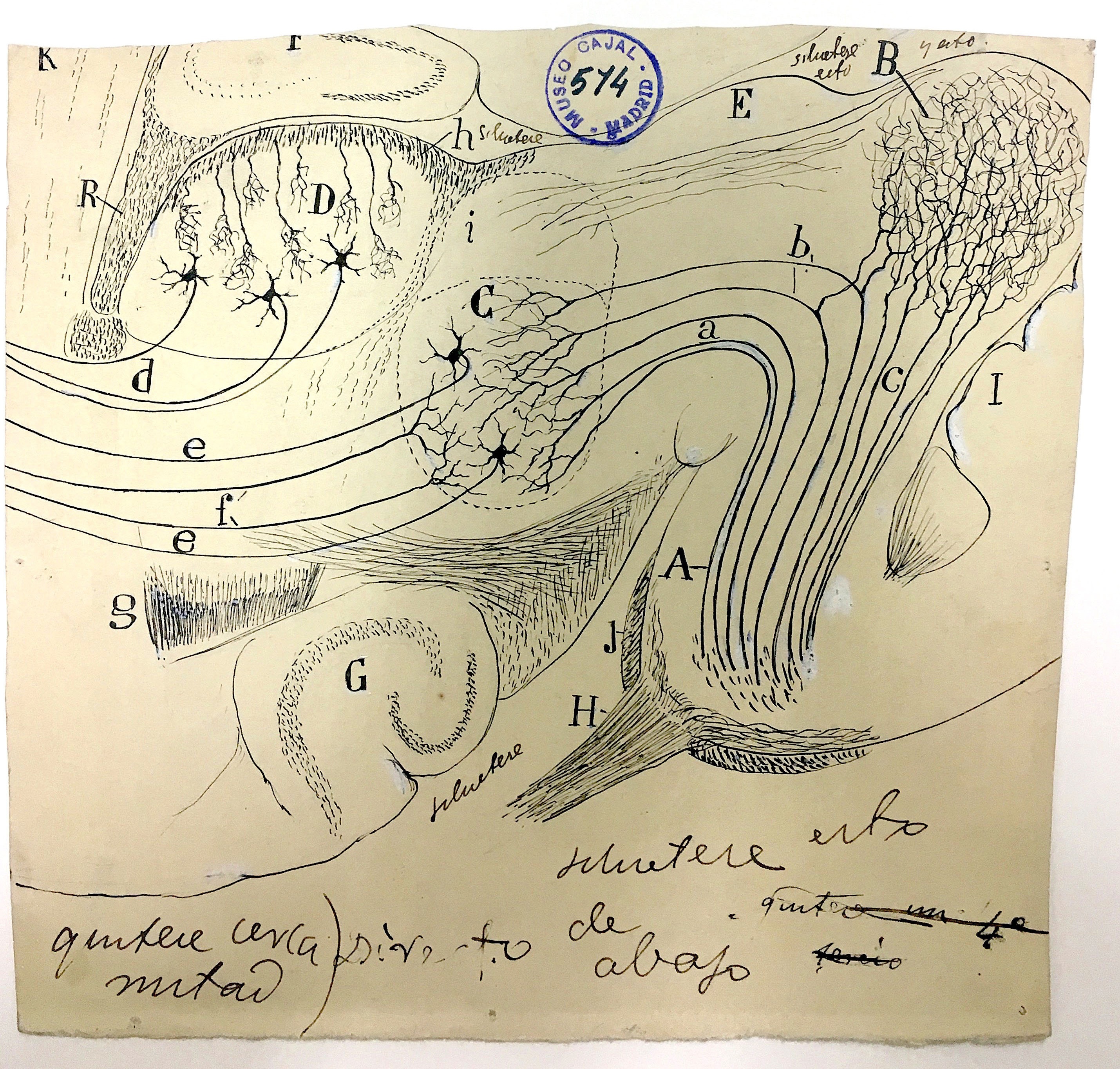
The mammalian auditory midbrain, shown above, is part of the ascending auditory pathway, responsible for relaying sensory signals from the ear into the primary auditory cortex deep in the brain. Cajal’s microscopy studies led him to believe that the lateral leminiscus (A) received input from the cochlear and superior olivary nuclei, and carried some of it to the inferior colliculus (B), which integrated the signals necessary for auditory reflexes, while the bulk of the information was sent directly to the medial geniculate body (C), which then relayed the information on to the auditory cortex via the thalamo-cortical path (e).
Modern studies have shown, however, that the inferior colliculus actually processes nearly all the input sent to the medial geniculate body and receives signals from the descending auditory pathway, as well as providing the motor integration necessary for auditory reflexes hypothesized by Cajal, making it a true hub for auditory signaling.
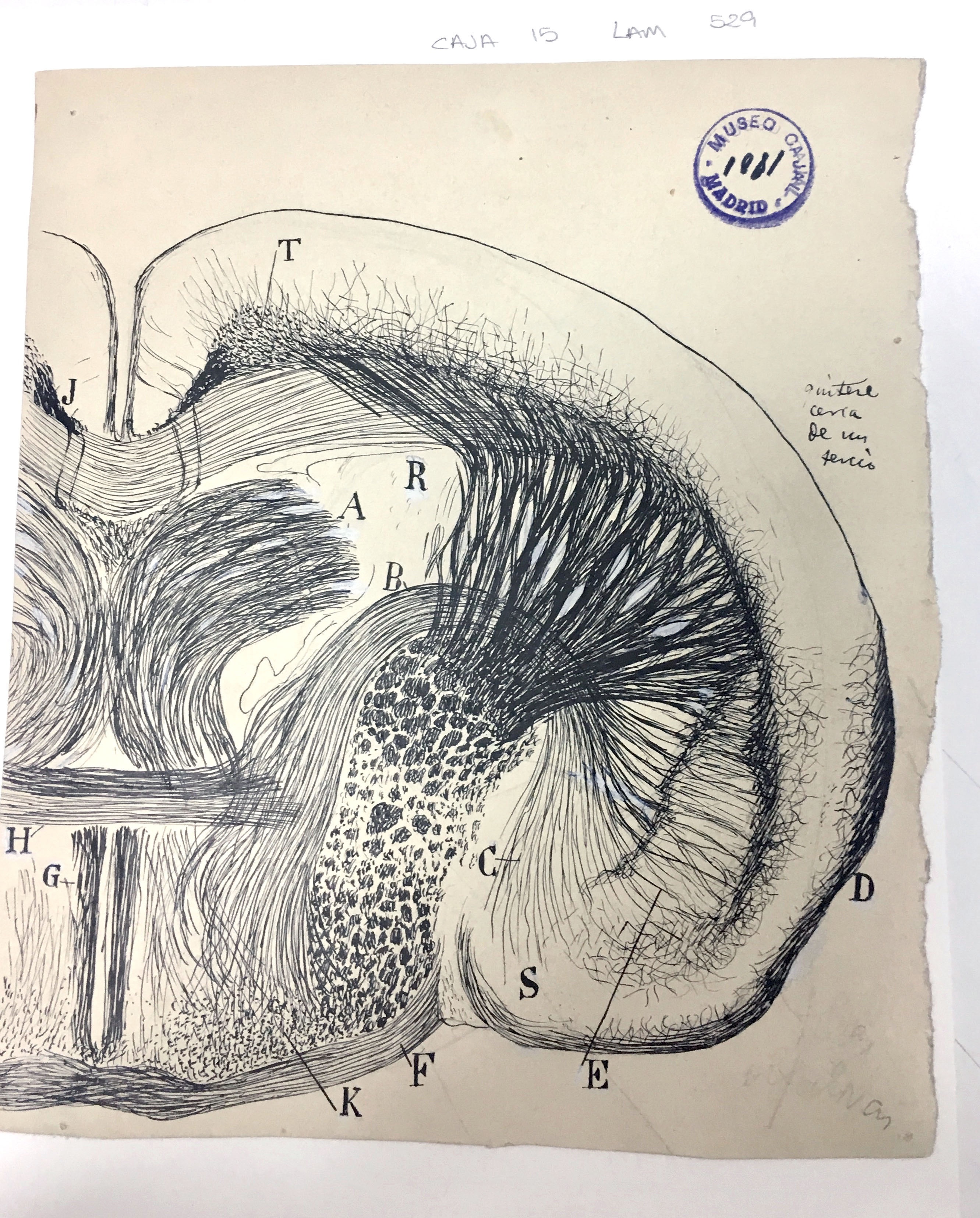
The lenticular nucleus (E) is a lens-shaped bundle of neurons that, along with the caudate nucleus (R) and the internal capsule, comprises the corpus striatum. Cajal used this drawing in his Texture of the Nervous System of Man and Vertebrates to illustrate the relatively large size of the lenticular nucleus in small mammals – in this case, a mouse – as compared to humans.
Although Cajal posited that the corpus striatum in general was of decreasing evolutionary importance and only useful for the coordination of higher reflexes, we now know that it is important for the facilitation of voluntary movement. The complexity and attention to detail in this drawing showcase Cajal’s skill in translating the view through his microscope lens to the page, where the structures he depicts are easily identifiable to today’s scientists more than 100 years after he put ink to paper.