
...
| Div | ||||||||||
|---|---|---|---|---|---|---|---|---|---|---|
| ||||||||||
|

...
4th Installation (current)
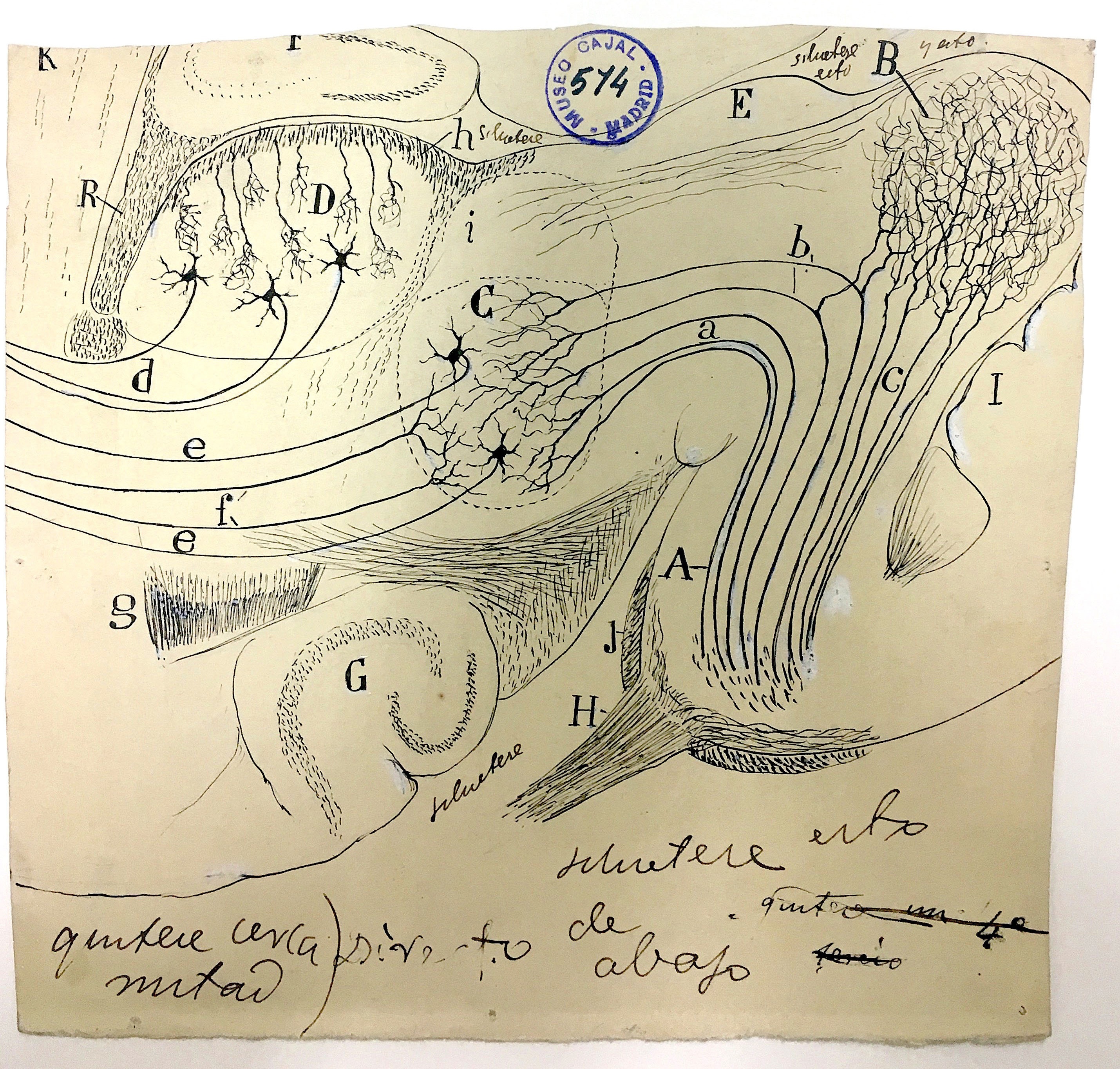
Nuclei in the auditory pathway
| Div | |||||||||||||||||||||
|---|---|---|---|---|---|---|---|---|---|---|---|---|---|---|---|---|---|---|---|---|---|
| |||||||||||||||||||||
|
Coronal brain section of a young mouse
| Div | |||||
|---|---|---|---|---|---|
| |||||
|
Coronal brain section of a young mouse
| |||||||||||||||||
| Div | |||||||||||||||||
|---|---|---|---|---|---|---|---|---|---|---|---|---|---|---|---|---|---|
| class | usa-grid
|
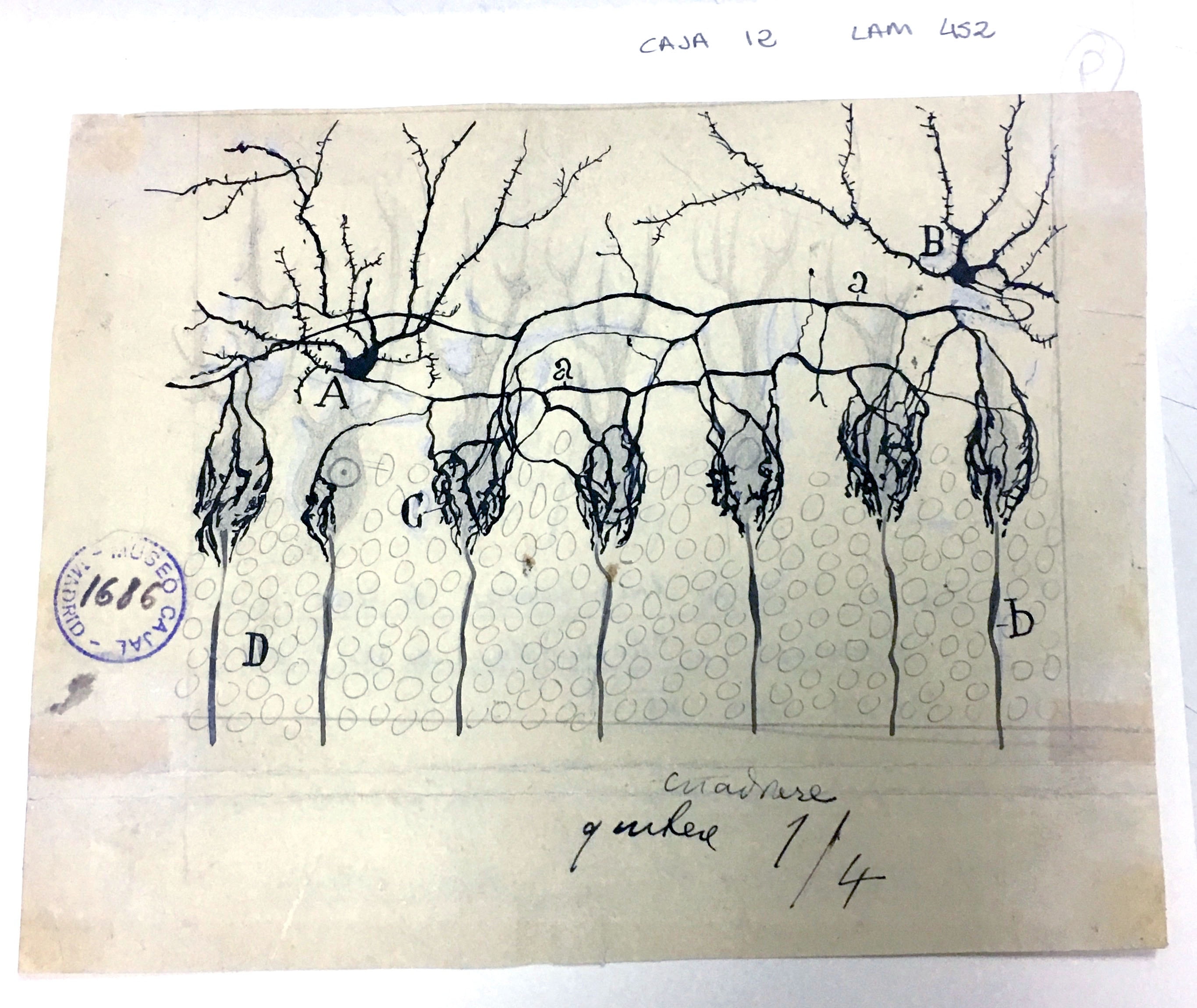
Basket cells in the cerebellum
| Div | |||||||||||||||
|---|---|---|---|---|---|---|---|---|---|---|---|---|---|---|---|
| |||||||||||||||
|
...
| Div | |||||||||||||||||||||
|---|---|---|---|---|---|---|---|---|---|---|---|---|---|---|---|---|---|---|---|---|---|
| |||||||||||||||||||||
|

Layer 5 of the visual cortex
| Div | ||||||||||||||||||||
|---|---|---|---|---|---|---|---|---|---|---|---|---|---|---|---|---|---|---|---|---|
| ||||||||||||||||||||
|

Interneurons of the auditory cortex
| Div | |||||||||||||||
|---|---|---|---|---|---|---|---|---|---|---|---|---|---|---|---|
| |||||||||||||||
|

...
Overview
Content Tools
ThemeBuilder