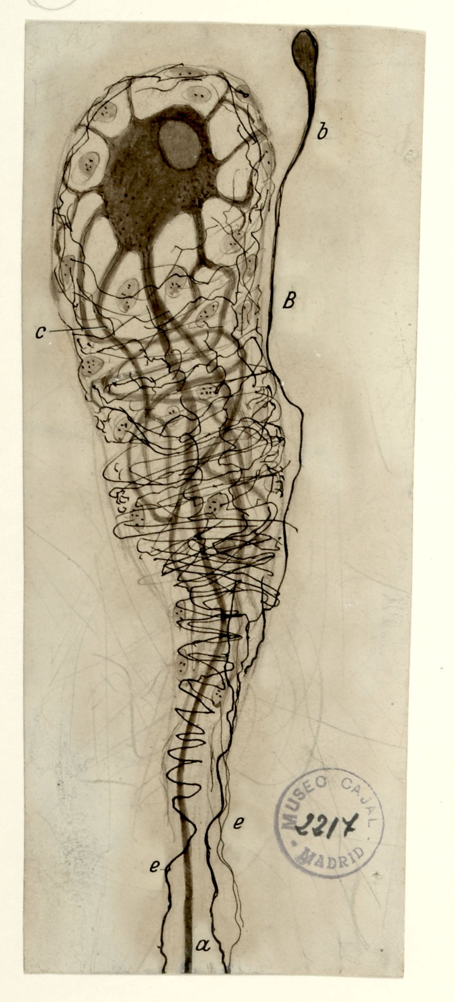
In 1905, Cajal studied human sympathetic ganglia and found morphological arrangements he had not yet seen in other species. Sympathetic ganglia comprises the thousands of afferent and efferent nerve cell bodies that run along either side of the spinal cord, connecting major organ systems, such as the renal system, to the spinal cord and brain. The kidney, a main organ within the renal system, filters blood to remove toxins via millions of structures called glomeruli, consisting of a tuft of blood vessels surrounded by a cuplike cellular structure known as Bowman’s Capsule. Shown above is a single neuron innervating a single glomerulus from a 50-year-old human subject, with a distinctive “comet” shape comprised of a very rich periglomerular nerve arborization. The kidney is innervated by both sympathetic and parasympathetic fibers; the innervating parasympathetic fibers originate from the vagal nerve. Pain signals caused by kidney stones may activate cross signaling along the vagal route, causing the well-known nausea and vomiting associated with kidney stones via the pathway shown in drawing no. 5.