
...
| Dive | |||||||||||||||||||||||||||||
|---|---|---|---|---|---|---|---|---|---|---|---|---|---|---|---|---|---|---|---|---|---|---|---|---|---|---|---|---|---|
| |||||||||||||||||||||||||||||
|

...
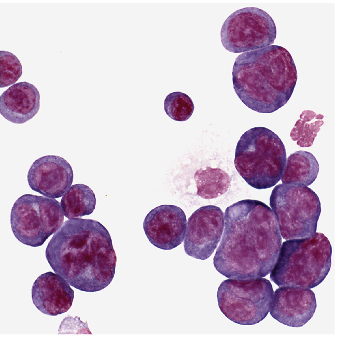
Potter was usually at his laboratory bench using this microscope. Shown are plasma cells with darkly stained nuclei. The clear spots next to the nucleus are perinuclear “hoffs” and are filled with newly synthesized protein, in this case, antibody.
Retroviruses Oncogene Activation
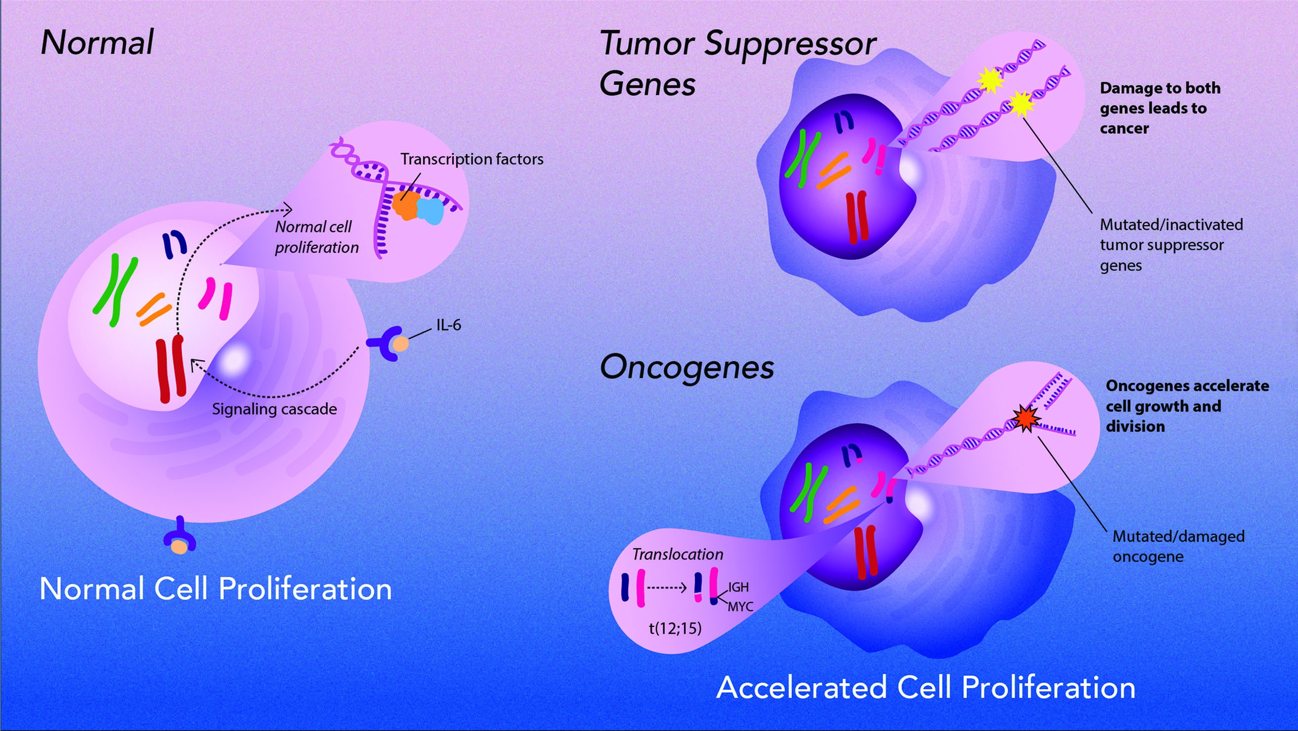
Potter collaborated with Marshall Sklar and Wallace Rowe of the National Institute of Allergy and Infectious Diseases to see if viruses could affect the growth of Potter’s plasma cell tumors. Although viruses did not cause the tumors, they found that retroviruses containing oncogenes could accelerate tumor growth when injected into mice, and could even induce tumors in normally resistant mice. Grace Shen-Ong, who worked in Potter’s laboratory, and Michael Cole then found that plasma cells that develop tumors have chromosomal translocations involving genes for immunoglobulins and an oncogene called MYC. But Potter — working with J. Frederic Mushinski, Ken Marcu, Rex Risser, and Siegfried Janz — found that these translocations alone did not cause tumors. And retroviruses had to contain both MYC and another oncogene to induce plasma cell tumors.
| Span | ||
|---|---|---|
| ||
Illustration designed by Sayeh Gorjifard, NCI, OD, CCT |
| Dive | ||||||||||||||||||||||||
|---|---|---|---|---|---|---|---|---|---|---|---|---|---|---|---|---|---|---|---|---|---|---|---|---|
| ||||||||||||||||||||||||
|

Overview
Content Tools
ThemeBuilder