Versions Compared

Key

  • This line was added.
  • This line was removed.
  • Formatting was changed.

...

Div
classgrid-row grid-gap
Div
classdesktop:grid-col-6:desktop

Each featured original illustration from the early 1900s, is accompanied by a caption written to engage scientists, researchers and investigators who populate the NIH campus.  As well as a 3-D printed rendering that enlarges a detail of the illustration above.  In this way, the drawings are rendered more accessible to a variety of audiences—including vision-impaired visitors who can directly experience these tactile versions of Cajal's drawings.  These files are made available on the 3D Print Exchange.  Direct links to the 3-D print files are provided at the end of this page.

The Cajal illustrations currently on-view include:

  • Auditory Tracts
  • Axonal Tracts in Rat
  • Cajal-Astrocytes
  • Cajal Astrocytes
  • Cerebral Cortex-ii
  • Interneuronal Plexuses
  • Medulla


Div
classdesktop:grid-col-6:desktop

Close up of Santiago Ramón y Cajal Exhibit illustration and 3d print

Div
classhr-3


Video

Widget Connector
width100%
urlhttps://www.youtube.com/watch?v=QIDUKHXqtg4
height560

Div
classhr-3


5th Installation (current)

Upper cervical ganglia

Div
classgrid-row grid-gap
Div
classdesktop:grid-col-6
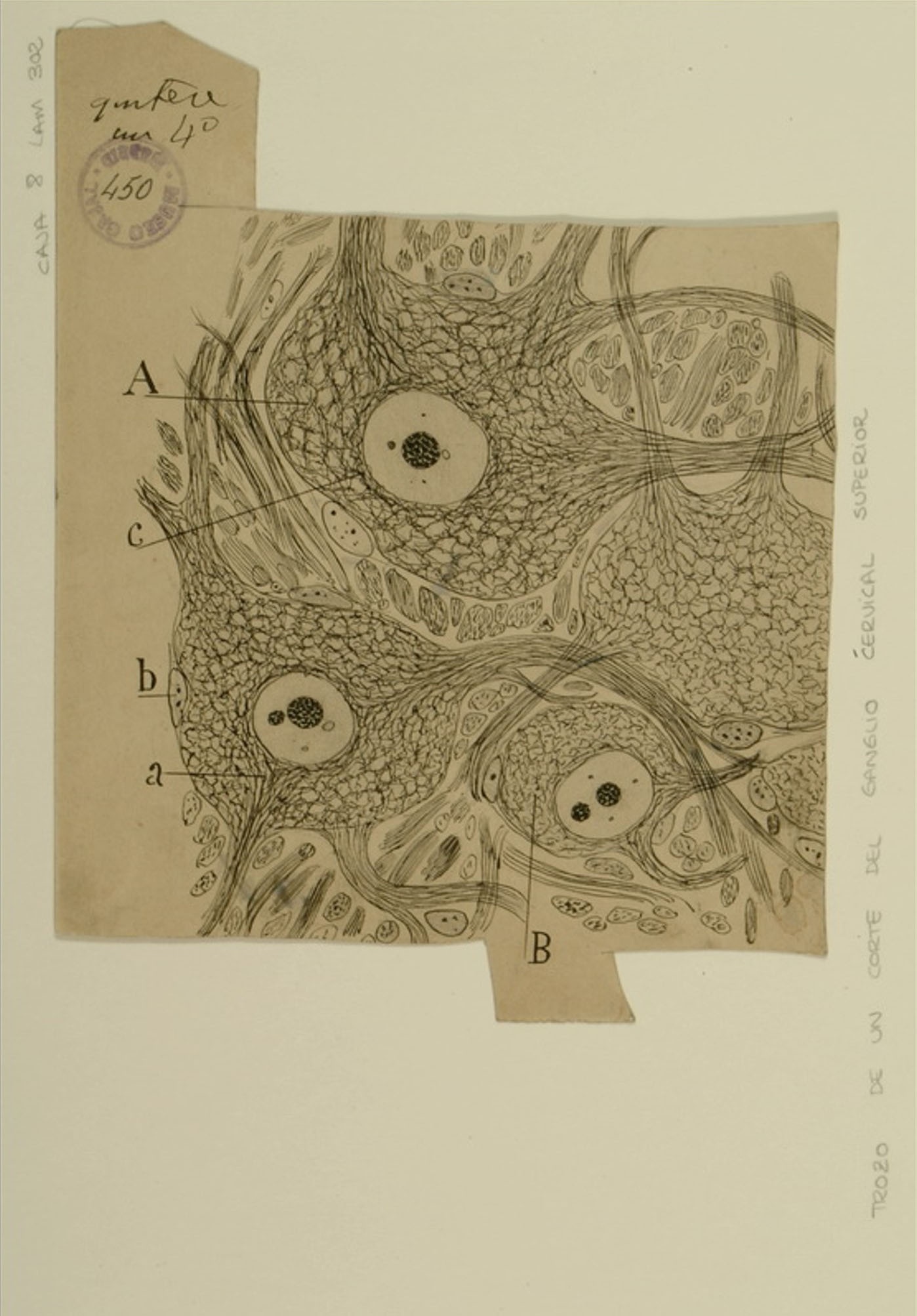
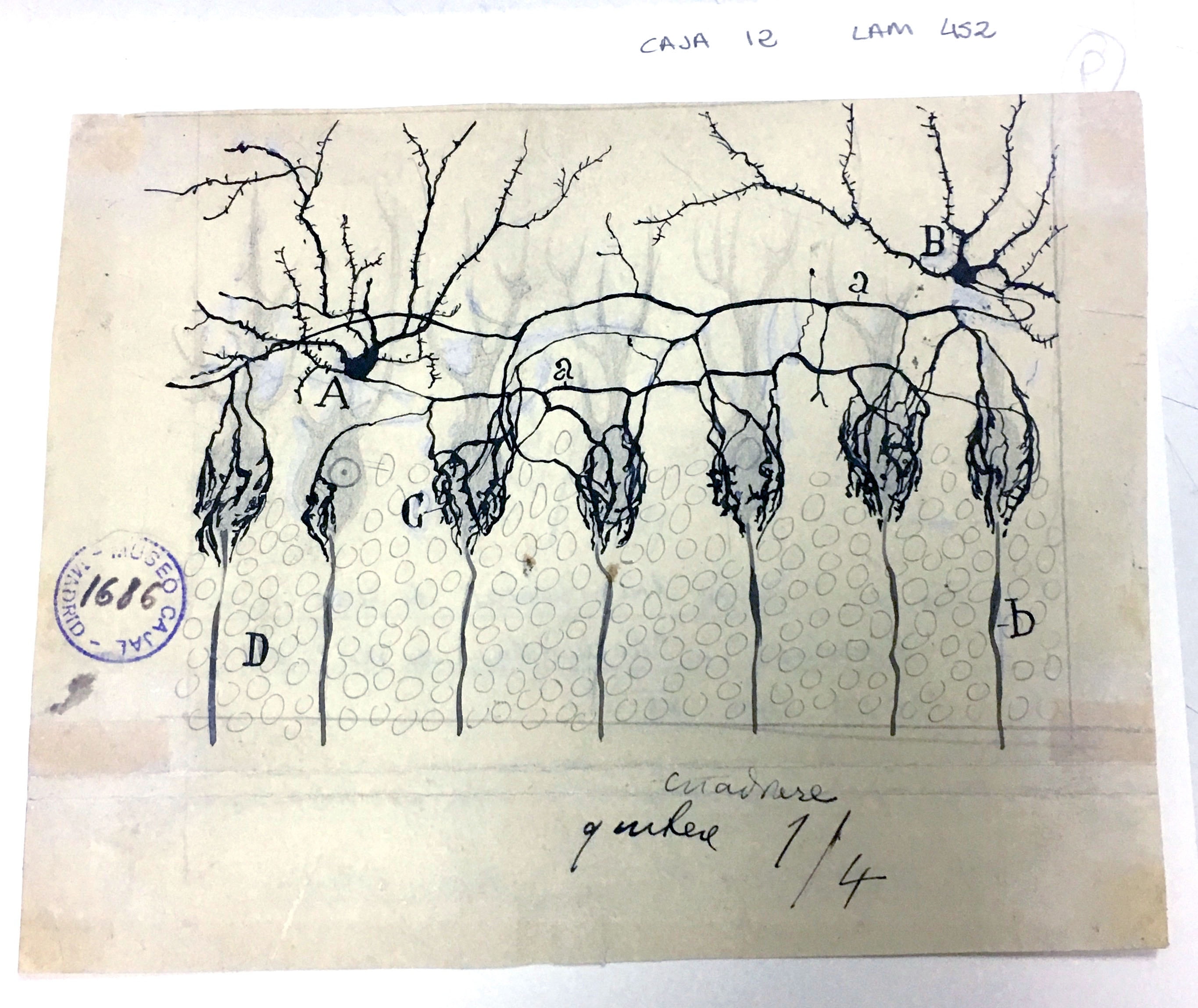
The autonomic nervous system controls involuntary body functions such as heart rate, digestion, and respiration, and is divided into two parts: the sympathetic nervous system and the parasympathetic nervous system. The sympathetic nervous system’s primary function is to activate the fight-or-flight response to danger, but it is also active at a basal level to preserve homeostasis. The superior cervical ganglion, a section of which is shown here, is a part of the sympathetic nervous system in which neurons originating in the spinal cord form synapses with neurons that innervate the heart, head, and neck, and control responses such as heart rate and pupil dilation. The cell bodies shown above are surrounded by “receptive nets” (A, B) formed by their own dendrites and the axons of neurons originating in the thoracic spinal cord. While Cajal himself performed preliminary work on the sympathetic nervous system, his former student, Fernando de Castro, gained international recognition for his careful studies of the fine structure of the autonomic ganglia. Cajal recognized de Castro as an equal and entrusted him with supervising the technical training and research of fellows at the Cajal Institute between 1924-1932.


Span
classusa-button

3dpx-010423 - Auditory Tracts

Div
classdesktop:grid-col-6

Span
classcredit

Courtesy of the Cajal Institute, Spanish National Research Council or CSIC©

Div
classhr-3


Comparison of visual and olfactory systems

Div
classgrid-row grid-gap
Div
classdesktop:grid-col-6

Cajal’s law of dynamic polarization, wherein information flows from the receptive dendrites of a neuron through its cell body and down through its axon (and on through the dendrites of the cells with which it forms synapses), is well illustrated in this figure above comparing the visual and olfactory systems. The olfactory and visual systems were integral to Cajal’s formulation of the law of dynamic polarization, since it was clear where the external stimulus originated and in which direction information ought to flow. Cajal was thus able to hypothesize that the dendrites (or, in photoreceptors, the pigmented outer segments) of the receptor neurons, which were closest to the stimulus, serve a receptive function, and thus, that the axons must be responsible for passing the information onwards. The olfactory system is shown in Fig 1: information originates in the olfactory mucosa (B), where olfactory receptor neurons (a) sense environmental stimuli and send signals to the olfactory bulb (A), which sends it further on to the olfactory cortex (C). This information transfer is unidirectional – signals originating in the olfactory cortex and relayed back to the olfactory bulb travel along a separate set of neurons (e,f,g). Similarly, in the visual system (Fig 2), information travels in the retina from the photoreceptors (a,m), through the bipolar cells (b,n) and ganglion cells (c,o) through the optic nerve to higher visual centers (B). Cajal showed that, in birds, information originating higher in the visual system travels back to the retina along a separate set of neurons (p,q,r).


Span
classusa-button

3dpx-010423 - Auditory Tracts

Div
classdesktop:grid-col-6

Span
classcredit

Courtesy of the Cajal Institute, Spanish National Research Council or CSIC©

Div
classhr-3


Medulla oblongata from a hemiplegic individual

Div
classgrid-row grid-gap
Div
classdesktop:grid-col-6
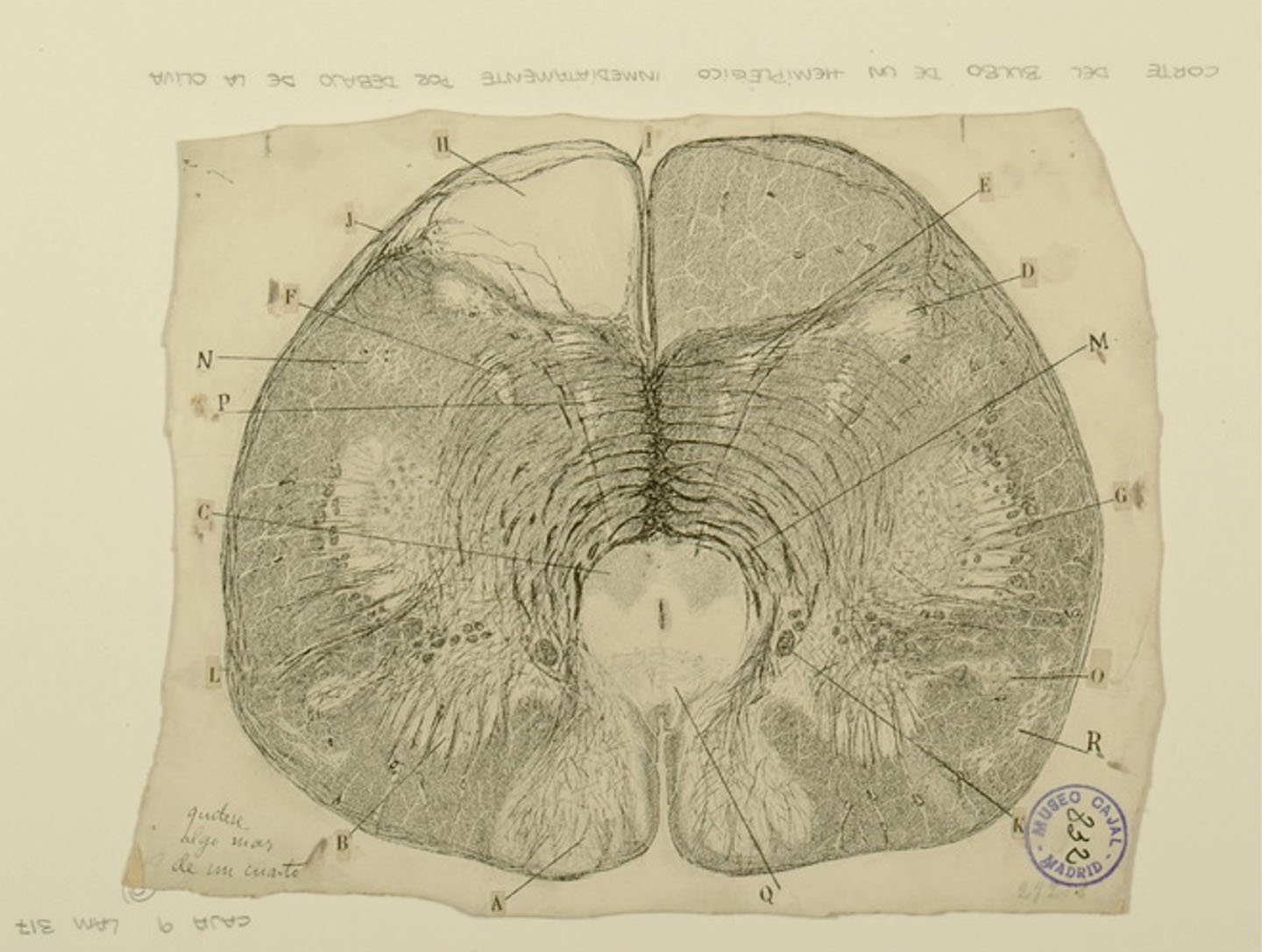
The medulla oblongata is a structure in the brainstem that controls involuntary bodily functions like respiration and heartbeat. Within the medulla, structures called medullary pyramids contain bundles of motor neuron fibers passing from the one side of the brain to the other side of the spinal cord. Injuries to the medullary pyramids may cause hemiplegia, or weakness/paralysis of one side of the body. In the figure above, a section of the medulla taken from an individual with hemiplegia, Cajal observes that the medullary pyramids on one side of the structure (H) have noticeably degenerated, leaving the space empty of neurons, while its counterpart on the other side remains intact. Other structures, such as the nucleus gracilis (A), and the nucleus cuneatus (B), which contain neurons responsible for fine touch and proprioception, appear to be undamaged. Cajal’s research at the time suggested there was little plasticity in the adult nervous system, and that treatment for hemiplegia was not possible; we now know that various types of therapies involving continued use and strengthening of the hemiplegic side of the body can promote at least small degrees of recovery.


Span
classusa-button

3dpx-010423 - Auditory Tracts

Div
classdesktop:grid-col-6

Span
classcredit

Courtesy of the Cajal Institute, Spanish National Research Council or CSIC©

Div
classhr-3


Innervation of Purkinje Cells

Div
classgrid-row grid-gap
Div
classdesktop:grid-col-6

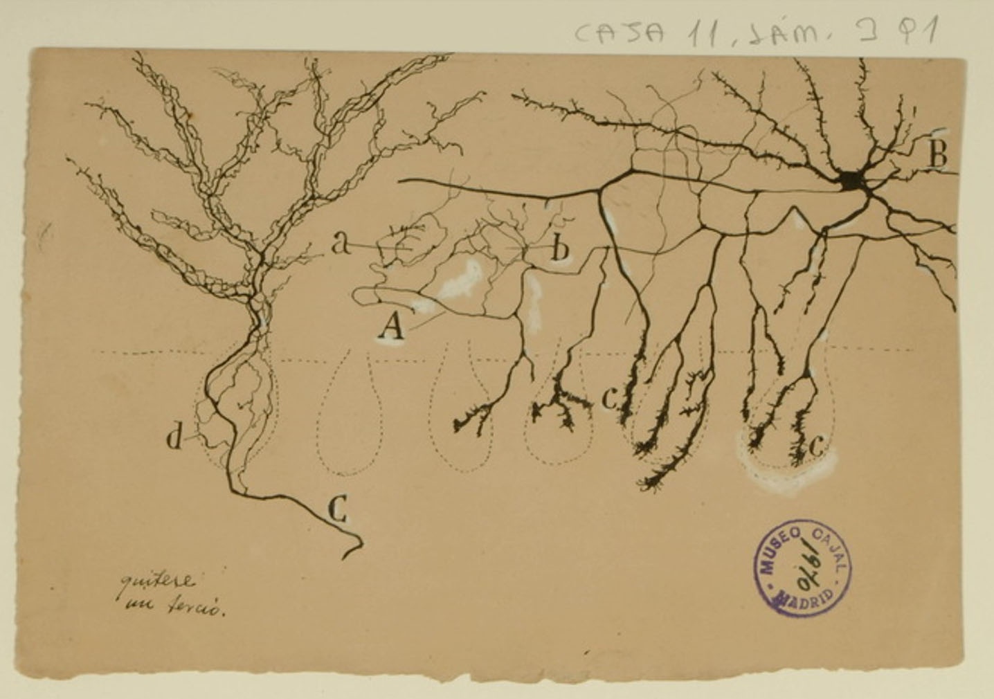
Purkinje cells are the main source of cerebellar output, and as such, they must be tightly controlled to correctly regulate motor functions. Basket cells regulate the output of Purkinje cells through the Pinceau formation: fine, brush-like basket cell axons in contact with Purkinje cell bodies (shown above in c; the Purkinje cell bodies are indicated by the faint dotted lines). Cajal described them in 1888, when it was yet unproven that neurons were separate cells rather than an interconnected reticulum, and correctly noted that the basket cells and Purkinje cells were distinct entities. He also correctly noted that the Pinceau formation was an exception to his law of dynamic polarization, where information flows from the axons of one neuron to the dendrites of a neighboring neuron: the axons of both the basket cells and the Purkinje cells interact in the Pinceau formation. He did not, however, foresee the biological importance of the Pinceau formation, which we now know to be critical for normal motor function.


Span
classusa-button

3dpx-010423 - Auditory Tracts

Div
classdesktop:grid-col-6

Span
classcredit

Courtesy of the Cajal Institute, Spanish National Research Council or CSIC©

Div
classhr-3


Mouse olfactory bulb

Div
classgrid-row grid-gap
Div
classdesktop:grid-col-6

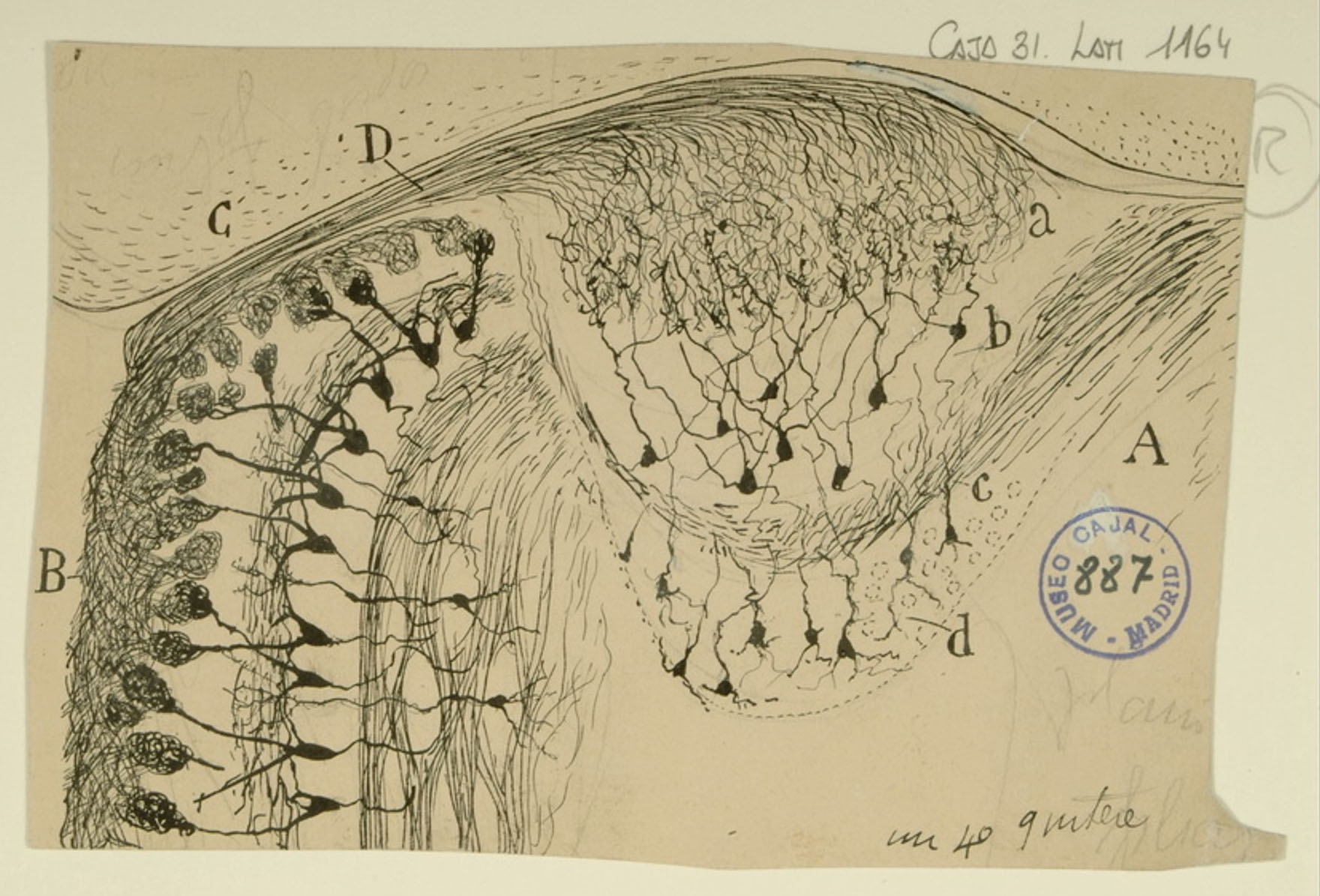
Cajal often used the olfactory system as a subject, finding it to be both easily accessible and regularly structured. Information derived from environmental stimuli that interact directly with olfactory receptors is transmitted to the main olfactory bulb (MOB) or accessory olfactory bulb (AOB) for processing before being transmitted deeper into the olfactory cortex. The MOB (B) receives input from the olfactory sensory neurons in the main olfactory epithelium, while the AOB (A) receives input from the vomeronasal organ via the vomeronasal nerve bundle (D). The AOB is thought to process input mainly from pheromones. Cajal noted that the AOB was composed of 4 layers: the glomerular layer (a), the mitral/tufted layer (b), the lateral olfactory tract (c), and the granular layer (d). Although the MOB and AOB shown above appear to be converging onto a common point, this is not the case – the mitral/tufted cells of the MOB synapse project to the main olfactory cortex, while the AOB mitral/tufted cells bypass the main olfactory cortex and project directly to the medial amygdala and hypothalamic nuclei.


Span
classusa-button

3dpx-010423 - Auditory Tracts

Div
classdesktop:grid-col-6

Span
classcredit

Courtesy of the Cajal Institute, Spanish National Research Council or CSIC©

Div
classhr-3


Mammalian retina

Div
classgrid-row grid-gap
Div
classdesktop:grid-col-6

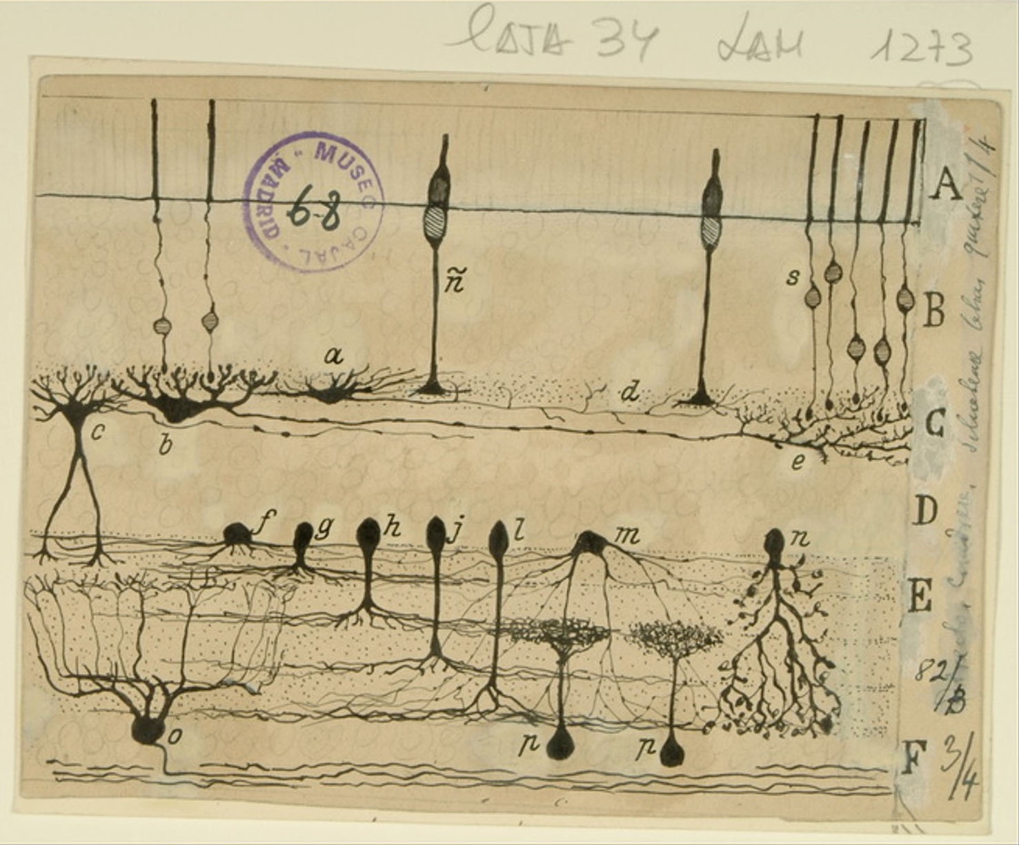
Cajal’s fascination with the retina is well documented – his publications on retinal neurons and organization in various species span 45 years, the length of his career. While many of Cajal’s representations of the retina focus on the visual pathway and the directionality of signal flow, this figure depicting a cross section of the mammalian retina showcases the wide variety of cell types therein, particularly amacrine cells (f-n). While the direction of information flow was clear in photoreceptors (ñ and s), bipolar cells (not pictured here) and ganglion cells (o), amacrine cells and horizontal cells (a,b) seemed to defy his law of dynamic polarization. Cajal imagined that the retina transmitted visual information like a complete photograph up into the visual cortex. We now know that the retina actually transmits many photographs in parallel, each depicting specific qualities of the visual world. Horizontal and amacrine cells shape the spatial and temporal characteristics of these parallel images, enabling ganglion cells to encode complex information about color, contrast, motion and other visual features.


Span
classusa-button

3dpx-010423 - Auditory Tracts

Div
classdesktop:grid-col-6

Span
classcredit

Courtesy of the Cajal Institute, Spanish National Research Council or CSIC©


...

Div
classgrid-row grid-gap
Div
classdesktop:grid-col-6
Basket cells are inhibitory interneurons found in several parts of the brain.  Those shown here, in the cerebellum, make motor movement possible by preventing inhibitory signaling from Purkinje neurons.  Each basket cell is composed of Purkinje neuron cell bodies surrounded by basketlike networks of axon branches (c) from the nearby stellate neurons (A and B); Cajal called these basketlike cell terminals ‘pinceau,’ French for ‘paintbrush.’  

Using the silver nitrate staining method to visualize these cells, he recognized that although the axons of the stellate neurons made numerous synapses with the Purkinje neuron cell bodies, they did not fuse at any point.  This supported his Neuron Doctrine, wherein the nervous system is composed of distinct cells rather than a network of continuously connected cells, and nervous impulses travel from the axon of one cell to the body of another.  

Although he first posited the Neuron Doctrine in 1894, it was not until the 1950s, when the first electron microscopes became available, that scientists were able to confirm the existence of the synapse and thus validate Cajal’s theory.


Span
classusa-button

3dpx-010425 - Cajal-Astrocytes

Div
classdesktop:grid-col-6

Span
classcredit

Courtesy of the Cajal Institute, Spanish National Research Council or CSIC©


Div
classhr-3


Astrocytes at the border of a wound

Div
classgrid-row grid-gap
Div
classdesktop:grid-col-6

Astrocytes are a type of macroglia that are critical for maintaining physiological homeostasis in the CNS and supporting neuronal function.  Astrocytes in the grey and white matter of the brain typically have pedicles, or “feet”, that form contacts with capillaries (A, B, e) and control local blood flow.  

Using a uranium-nitrate technique specifically for staining astrocytes on a tissue sample bordering a cerebral wound, Cajal observed not only normal astrocytes in contact with capillaries, but also small amoeboid cells (a,b,c).  Other scientists, such as Alzheimer, had previously noted such cells in the CNS tissue of persons with various degenerative diseases, but their origins were uncertain.  Cajal correctly inferred that these cells were astrocytes which had somehow reshaped themselves after the injury.  

We now know that astrocytes become “reactive” after a brain injury: they become polarized, migrate, and their cell bodies swell.  Such reactive astrocytes are postulated to have both beneficial (wound healing, limitation of inflammation) and detrimental (scar formation) roles in the response to injury.


Span
classusa-button

3dpx-010426 - Cajal-Astr

Div
classdesktop:grid-col-6

Span
classcredit

Courtesy of the Cajal Institute, Spanish National Research Council or CSIC©


...