Versions Compared

Key

  • This line was added.
  • This line was removed.
  • Formatting was changed.

...

Div
classgrid-row grid-gap
Div
classgrid-col-6

On March 31, 2014, the second phase of the John Edward Porter Neuroscience Research Center was dedicated.  This new facility is shared by scientists from the National Institute of Neurological Disorders and Stroke (NINDS), National Eye Institute (NEI), National institute of Child Health and Human Development (NICHD), National Institute of Dental and Craniofacial Research (NIDCR), National institute of Mental Health (NIMH), National Institute on Deafness and Other Communicable Disorders (NIDCD), and the National Institute of Biomedical Imaging and Bioengineering (NBIB)—and represents a unique opportunity for scientists to collaborate across academic disciplines as well as the boundaries that sometimes separate institutes and centers on the NIH campus.  

A new wave of research and exploration is beginning within these walls with new support for the creation of a new arsenal of instruments for unlocking the mysteries of the brain through the Brain Research through Advancing Innovative Neurotechnologies (BRAIN) Initiative.  This moment may be the most appropriate to look back over the accomplishments of the last century and anticipate those of the next. 

Table of Contents
maxLevel2
minLevel2

Div
classgrid-col-6

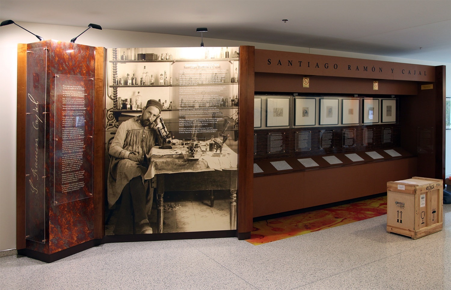
Santiago Ramón y Cajal exhibit with shipping crate

Center
Span
classcaption

This Exhibit currently located in building 35

Div
classhr-3


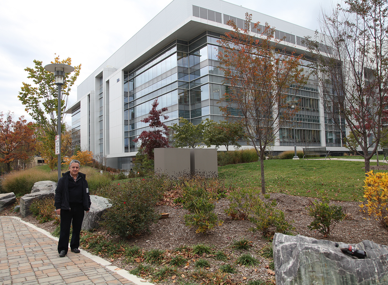
Ricardo Martínez Murillo, Ph.D., Director of the Instituto Cajal in Madrid, Spain, is pictured in front of NIH's recently dedicated 
neuroscience research center where the exhibition of Ramón y Cajal original drawings is located.

...

Div
classgrid-row grid-gap
Div
classgrid-col-6

Cajal’s law of dynamic polarization, wherein information flows from the receptive dendrites of a neuron through its cell body and down through its axon (and on through the dendrites of the cells with which it forms synapses), is well illustrated in this figure above comparing the visual and olfactory systems. The olfactory and visual systems were integral to Cajal’s formulation of the law of dynamic polarization, since it was clear where the external stimulus originated and in which direction information ought to flow. Cajal was thus able to hypothesize that the dendrites (or, in photoreceptors, the pigmented outer segments) of the receptor neurons, which were closest to the stimulus, serve a receptive function, and thus, that the axons must be responsible for passing the information onwards. The olfactory system is shown in Fig 1: information originates in the olfactory mucosa (B), where olfactory receptor neurons (a) sense environmental stimuli and send signals to the olfactory bulb (A), which sends it further on to the olfactory cortex (C). This information transfer is unidirectional – signals originating in the olfactory cortex and relayed back to the olfactory bulb travel along a separate set of neurons (e,f,g). Similarly, in the visual system (Fig 2), information travels in the retina from the photoreceptors (a,m), through the bipolar cells (b,n) and ganglion cells (c,o) through the optic nerve to higher visual centers (B). Cajal showed that, in birds, information originating higher in the visual system travels back to the retina along a separate set of neurons (p,q,r).


Span
classusa-button

3dpx-010423 - Auditory Tracts

Div
classgrid-col-6

Image Modified

Span
classcredit

Courtesy of the Cajal Institute, Spanish National Research Council or CSIC©

...

Div
classgrid-row grid-gap
Div
classgrid-col-6
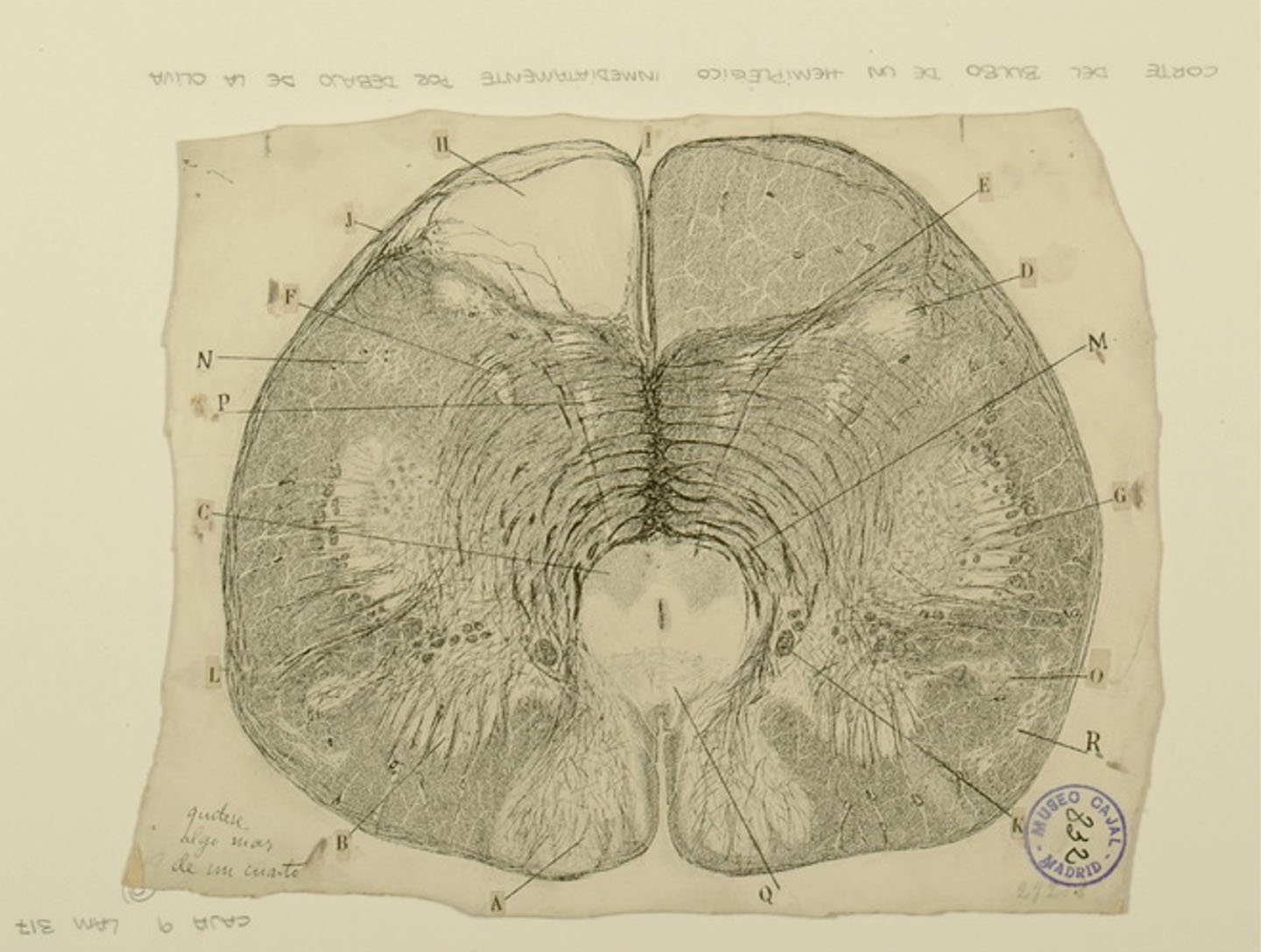
The medulla oblongata is a structure in the brainstem that controls involuntary bodily functions like respiration and heartbeat. Within the medulla, structures called medullary pyramids contain bundles of motor neuron fibers passing from the one side of the brain to the other side of the spinal cord. Injuries to the medullary pyramids may cause hemiplegia, or weakness/paralysis of one side of the body. In the figure above, a section of the medulla taken from an individual with hemiplegia, Cajal observes that the medullary pyramids on one side of the structure (H) have noticeably degenerated, leaving the space empty of neurons, while its counterpart on the other side remains intact. Other structures, such as the nucleus gracilis (A), and the nucleus cuneatus (B), which contain neurons responsible for fine touch and proprioception, appear to be undamaged. Cajal’s research at the time suggested there was little plasticity in the adult nervous system, and that treatment for hemiplegia was not possible; we now know that various types of therapies involving continued use and strengthening of the hemiplegic side of the body can promote at least small degrees of recovery.


Span
classusa-button

3dpx-010423 - Auditory Tracts

Div
classgrid-col-6

Image Modified

Span
classcredit

Courtesy of the Cajal Institute, Spanish National Research Council or CSIC©

...

Div
classgrid-row grid-gap
Div
classgrid-col-6

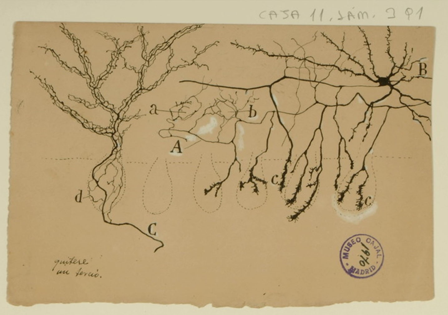
Purkinje cells are the main source of cerebellar output, and as such, they must be tightly controlled to correctly regulate motor functions. Basket cells regulate the output of Purkinje cells through the Pinceau formation: fine, brush-like basket cell axons in contact with Purkinje cell bodies (shown above in c; the Purkinje cell bodies are indicated by the faint dotted lines). Cajal described them in 1888, when it was yet unproven that neurons were separate cells rather than an interconnected reticulum, and correctly noted that the basket cells and Purkinje cells were distinct entities. He also correctly noted that the Pinceau formation was an exception to his law of dynamic polarization, where information flows from the axons of one neuron to the dendrites of a neighboring neuron: the axons of both the basket cells and the Purkinje cells interact in the Pinceau formation. He did not, however, foresee the biological importance of the Pinceau formation, which we now know to be critical for normal motor function.


Span
classusa-button

3dpx-010423 - Auditory Tracts

Div
classgrid-col-6

Image Modified

Span
classcredit

Courtesy of the Cajal Institute, Spanish National Research Council or CSIC©

...

Div
classgrid-row grid-gap
Div
classgrid-col-6

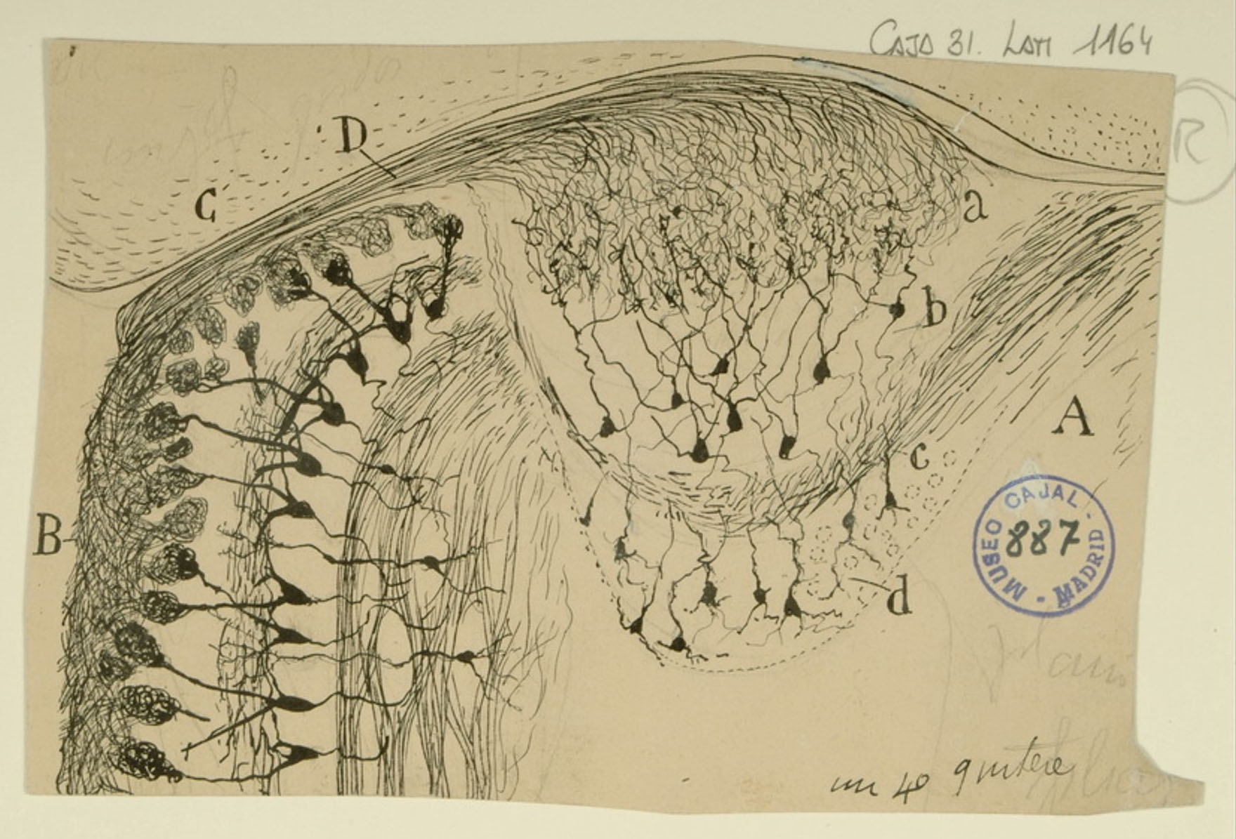
Cajal often used the olfactory system as a subject, finding it to be both easily accessible and regularly structured. Information derived from environmental stimuli that interact directly with olfactory receptors is transmitted to the main olfactory bulb (MOB) or accessory olfactory bulb (AOB) for processing before being transmitted deeper into the olfactory cortex. The MOB (B) receives input from the olfactory sensory neurons in the main olfactory epithelium, while the AOB (A) receives input from the vomeronasal organ via the vomeronasal nerve bundle (D). The AOB is thought to process input mainly from pheromones. Cajal noted that the AOB was composed of 4 layers: the glomerular layer (a), the mitral/tufted layer (b), the lateral olfactory tract (c), and the granular layer (d). Although the MOB and AOB shown above appear to be converging onto a common point, this is not the case – the mitral/tufted cells of the MOB synapse project to the main olfactory cortex, while the AOB mitral/tufted cells bypass the main olfactory cortex and project directly to the medial amygdala and hypothalamic nuclei.


Span
classusa-button

3dpx-010423 - Auditory Tracts

Div
classgrid-col-6

Image Modified

Span
classcredit

Courtesy of the Cajal Institute, Spanish National Research Council or CSIC©

...

Div
classgrid-row grid-gap
Div
classgrid-col-6

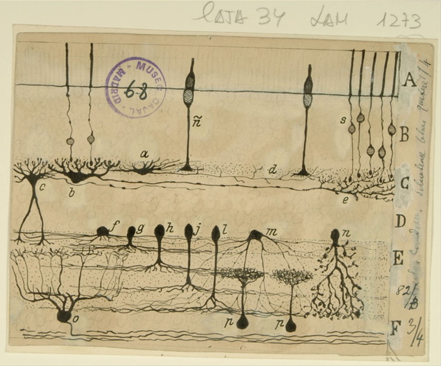
Cajal’s fascination with the retina is well documented – his publications on retinal neurons and organization in various species span 45 years, the length of his career. While many of Cajal’s representations of the retina focus on the visual pathway and the directionality of signal flow, this figure depicting a cross section of the mammalian retina showcases the wide variety of cell types therein, particularly amacrine cells (f-n). While the direction of information flow was clear in photoreceptors (ñ and s), bipolar cells (not pictured here) and ganglion cells (o), amacrine cells and horizontal cells (a,b) seemed to defy his law of dynamic polarization. Cajal imagined that the retina transmitted visual information like a complete photograph up into the visual cortex. We now know that the retina actually transmits many photographs in parallel, each depicting specific qualities of the visual world. Horizontal and amacrine cells shape the spatial and temporal characteristics of these parallel images, enabling ganglion cells to encode complex information about color, contrast, motion and other visual features.


Span
classusa-button

3dpx-010423 - Auditory Tracts

Div
classgrid-col-6

Image Modified

Span
classcredit

Courtesy of the Cajal Institute, Spanish National Research Council or CSIC©

...

Div
classgrid-row grid-gap
Div
classgrid-col-6

The superior temporal gyrus contains the auditory cortex, responsible for processing sound, and Wernicke’s area, which is necessary for the processing of speech to be understood as language rather than simply sounds. Shown here are several layers of pyramidal cells in the superior temporal gyrus, which is layered similarly to other areas of the temporal cortex. Though they vary in size and position, the pyramidal cells (a,b,c,d,e,f,g,h) all exhibit the characteristic cone-shaped cell body, a single apical dendrite extending upwards to the cortical surface, basal dendrites, and basal axons (a). Cajal characterized pyramidal cells from many tissues, detailing the variety of shapes and sizes found in different locations throughout the brain. He also hypothesized that size and shape of the dendritic arborizations of the pyramidal cells would vary over the lifespan of an organism depending; recent research reveals that the cells shown here, from the superior temporal gyrus of an infant, have much larger and denser dendritic branches than those from an adult would in this specific location.


Span
classusa-button

3dpx-010423 - Auditory Tracts

Div
classgrid-col-6

Image Modified

Span
classcredit

Courtesy of the Cajal Institute, Spanish National Research Council or CSIC©

...

Div
classgrid-row grid-gap
Div
classgrid-col-6

3d print


Center
Span
classcaption

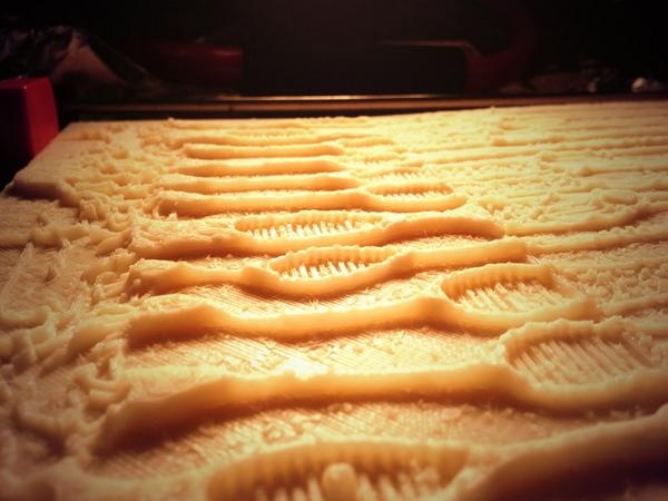
Close up of the 3D print

Div
classgrid-col-6

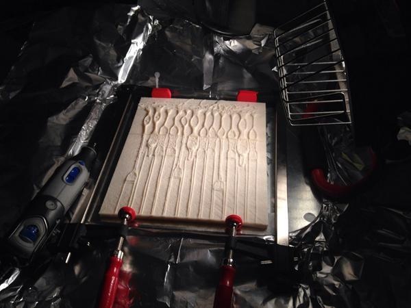
3D Print in a vice to hold it as the solvent dries

Center
Span
classcaption

Photograph of the 3D printed plates being bonded together with acetone

Acknowledgements

This exhibition is a collaborative effort of the Office of NIH History’s Stetten Museum and the National Institute of Neurological Disorders and Stroke (NINDS).  Jeffrey S. Diamond, Ph.D., Senior Investigator, Synaptic Physiology Section, Division of Intramural Research, NINDS, represented the NIH in negotiating the loan of the original drawings from the Cajal Institute of Madrid, Spain.  NIH and the exhibition team are particularly grateful to Dr. Juan De Carlos, of the Cajal Institute for making this exhibition possible through the loan of the original drawings, and the archival images featured in the exhibition. 

...