Versions Compared

Key

  • This line was added.
  • This line was removed.
  • Formatting was changed.

...

Div
classgrid-row grid-gap

What causes RMSF?


Div
classgrid-col-6

Collecting Ticks

Although it was known that RMSF was caused when the Rickettsia rickettsii bacteria infected a Dermacentor andersoni tick that then bit a human, more questions remained. Was the bacteria spread by other species of ticks? And exactly how did the ticks get infected? To find out where the ticks were picking up the infection and to answer a number of other questions, ticks had to be collected in the wild.

Div
classgrid-col-6

Image RemovedMan dressed in white with pants tucked in boots waves large white flag over meadow grass in high mountains.Image Added

A staff member of the Canyon Creek Schoolhouse Laboratory—possibly Dr. Ralph Parker, C.M. Salisbury, or George Cowan—dragged a white flannel flag over brush and grass to gather ticks in the Bitterroot Valley, Montana. He wore a white jump suit over his regular clothes, tucking his pant legs into the top of his high, laced boots. When they returned from such outings, the men would check each other closely in case a tick had attached itself to one of them.

Image: Office of NIH History and Stetten Museum, 1522

In 1923, a mountain goat (perhaps this mountain goat kid) taken by George Cowan had over 1,000 ticks engorged ticks on it. It was with these ticks that Dr. Roscoe Spencer came up with his idea for an RMSF vaccine.

Image: Montana Memory, 264

Div
classgrid-row grid-gap
Div
classgrid-col-6

Cataloguing Ticks

As the field researchers gathered many species of ticks and ticks in many stages of their life cycle, Dr. Robert Cooley developed a huge collection of them. Cooley was the head entomologist at Canyon Creek Schoolhouse laboratory. This collection would help later researchers solve questions about other tick-borne diseases.

Div
classgrid-col-6

Dr. Robert Cooley shows off his world-class tick collection. The second picture was taken in Building One (which opened in 1928), not the Canyon Creek Schoolhouse laboratory, on December 3, 1946. Cooley had been developing this collection since his first days studying RMSF forty years before.

[RML may have a photo like these in high res that could be used instead.]

Image: Wikimedia Commons 

...

Div
classgrid-row grid-gap
Div
classgrid-col-6

Life cycle of ticks

Studying the life cycle of ticks provided important pieces of the RMSF puzzle. Knowing the life cycle meant that scientists could study questions such as: When did the ticks pick up the bacteria causing RMSF—was it when they were newly hatched? When they were adults? And was the infection passed on to a new generation of ticks through the eggs? The answers to these questions could be used in control efforts and in developing a new vaccine.

Div
classgrid-col-6

Image RemovedTick with eggs bunched together in bundle larger than tickImage Added

The life cycle of the tick: a fertilized female lays thousands of eggs on the ground. When they hatch as larvae, they attach to small animals and feed. Then they drop off to digest their meal, and molt (shed their old shell) to become nymphs. The first winter they spend as unfed nymphs.

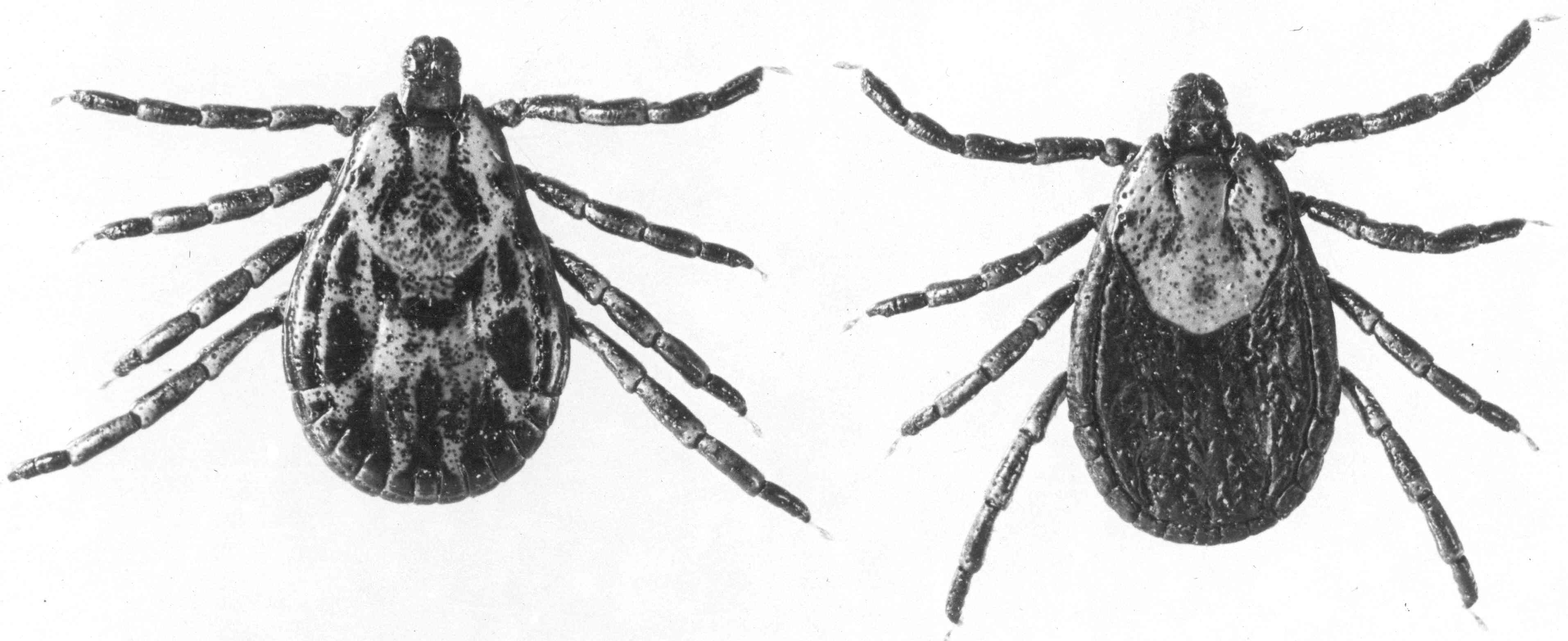
In spring, the nymphs once again attach to an animal and feed, drop to the ground, and digest their meals. This time they molt to adults, like these Rocky Mountain wood ticks, Dermacentor andersoni. They spend their second winter as unfed adults.

The next spring they climb grass and bushes to attach to animals.


But as adults, ticks mate as well as feed. This is when the ticks can get the Rickettsia rickettsii bacteria from the animals they feed on. Even though the bacteria don’t hurt the ticks, it infects all of their cells, and is passed on through the egg. The next generation of ticks will be infected.

Images: Office of NIH History and Stetten Museum, 1454-58

...