
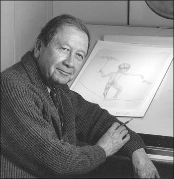
The Ultimate Portrait Painter
Howard Bartner and 40 Years of Medical Science
Introduction
| Div | ||||||||||||||||
|---|---|---|---|---|---|---|---|---|---|---|---|---|---|---|---|---|
| ||||||||||||||||
|
| Div | ||||||||||
|---|---|---|---|---|---|---|---|---|---|---|
| ||||||||||
|
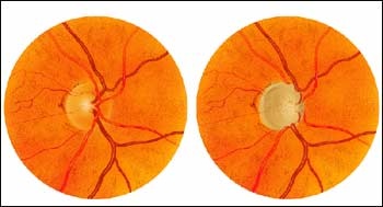
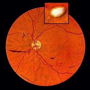
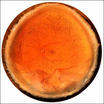
Ophthalmological Disorders
In this series of paintings of the inside of the eye, the orange circle represents the retina and the yellow circle represents the optic disc, which is where the optic nerve that connects the eye to the brain leaves the retina.
| Div | ||||||||||
|---|---|---|---|---|---|---|---|---|---|---|
| ||||||||||
|
| Div | ||||||||||
|---|---|---|---|---|---|---|---|---|---|---|
| ||||||||||
|
Anatomy of the Infant Head
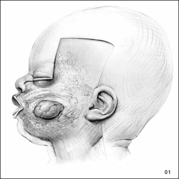
Because infant anatomy of the head differs greatly from adult cranial anatomy, Dr. James Bosma of the National Institute of Dental and Craniofacial Research wanted to produce a reference guide on infant anatomy for pediatricians, pediatric dentists, maxillofacial surgeons, and other physicians. Bosma hoped that by learning more about the development of the infant airway, throat, larynx, and pharynx, that doctors could better understand breast feeding and respiratory abnormalities. Bosma asked Howard Bartner to collaborate with him and three other illustrators in the study and description of the infant head, resulting in the publishing of Anatomy of the Infant Head (Johns Hopkins University Press, 1986). Bartner's elegant illustrations are important features of this classic text.
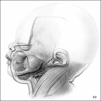
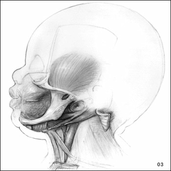
Below are side-view drawings of an infant's head. Note the nugget of extra fat, known as the "buccal pad." The buccal pad contributes to babies' chubby cheeks and diminishes when the infant is about a year old. Drawings two and three depict arrangements of the jaw and throat muscles.
| Div | |||||||||||||||
|---|---|---|---|---|---|---|---|---|---|---|---|---|---|---|---|
| |||||||||||||||
|
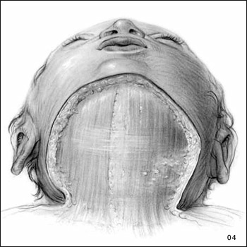
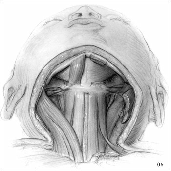
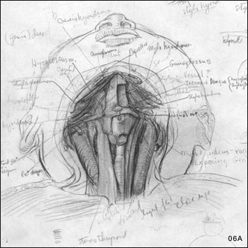
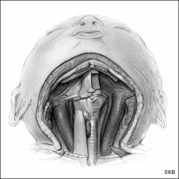
Sequential Views of the Infant's Throat, Larynx Areas, and Mouth Muscles from the Front
Removal of the skin shows tissue and muscles in the throat area. Note how the drawings become progressively more detailed as the levels of dissection go deeper. The left side of drawing 6 is a working sketch; the finished product is on the right.
| Div | ||||||||||
|---|---|---|---|---|---|---|---|---|---|---|
| ||||||||||
|
| Div | ||||||||||
|---|---|---|---|---|---|---|---|---|---|---|
| ||||||||||
|
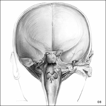
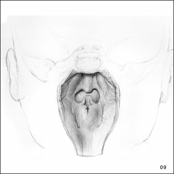
Drawings 7-9: Sequential Drawings of the Throat Area from the Back
The following drawings show the back of an infant's head. Drawing seven depicts the relation between the pharynx and vessels and nerves on each side of it. The eighth drawing illustrates the ear as seen from the back; and the ninth drawing is a close-up of the pharynx and larynx areas.
| Div | |||||||||||||||
|---|---|---|---|---|---|---|---|---|---|---|---|---|---|---|---|
| |||||||||||||||
|

Human Physiology in Space
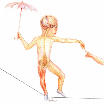
In some of his most recent work, Bartner created illustrations for the textbook Human Physiology in Space, produced jointly by the National Aeronautics and Space Administration (NASA), NIH, the Universities Space Research Association, and the University of Texas Southwestern Medical Center. The textbook uses space science to inspire high school students to learn about human physiology. Bartner invented two characters, a teenage boy and girl, to demonstrate the ideas in the text; his whimsical drawings keep students involved.
Drawing 1: Boy on a Tightrope
| Div | ||||||||||
|---|---|---|---|---|---|---|---|---|---|---|
| ||||||||||
|
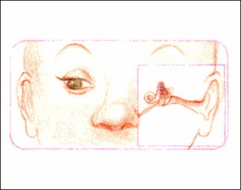
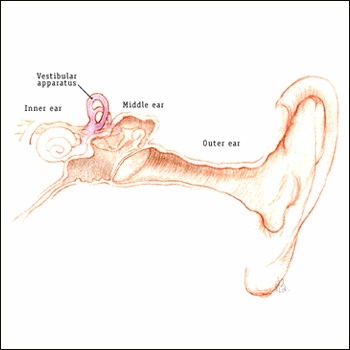
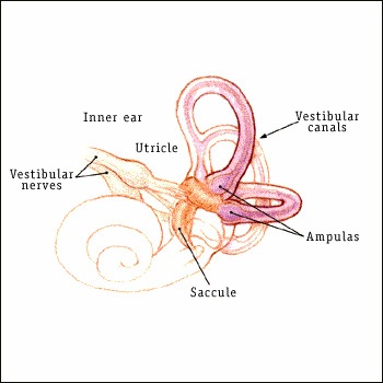
Drawing 2: The Inner Ear
In this series of drawings, Bartner shows in increasing detail the vestibule organs of the inner ear responsible for balance. The three curved canals of the inner ear detect when the head rotates, and the two small organs they join (the otolith organs) detect when the head moves linearly. That's why an infection in your inner ear may affect your balance.
| Div | |||||||||||||||
|---|---|---|---|---|---|---|---|---|---|---|---|---|---|---|---|
| |||||||||||||||
|
Photography Credits
All images, text, transcription, and/or recordings reproduced in these exhibits and galleries may be protected by copyright. Users who wish to reproduce materials for other than private use should make an additional effort to determine ownership and, if restricted, seek permission before reproducing them.
Related Links
- Johns Hopkins University Medical Arts Program
- National Eye Institute
- Glaucoma Research Foundation
- Johns Hopkins University Press
- National Institute of Child Health and Human Development
- National Institute of Dental and Craniofacial Research
- National Aeronautics and Space Administration
- Universities Space Research Association
- University of Texas Southwestern Medical Center
Overview
Content Tools
ThemeBuilder