
| Div | ||||||||||||||||
|---|---|---|---|---|---|---|---|---|---|---|---|---|---|---|---|---|
| ||||||||||||||||
|
| Div | ||
|---|---|---|
| ||
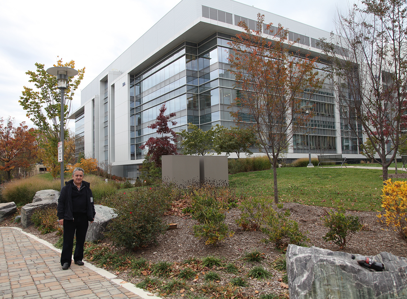 Ricardo Martínez Murillo, Ph.D., Director of the Instituto Cajal in Madrid, Spain, is pictured in front of NIH's recently dedicated
neuroscience research center where the exhibition of Ramón y Cajal original drawings is located.
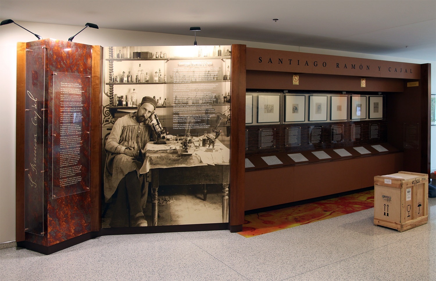
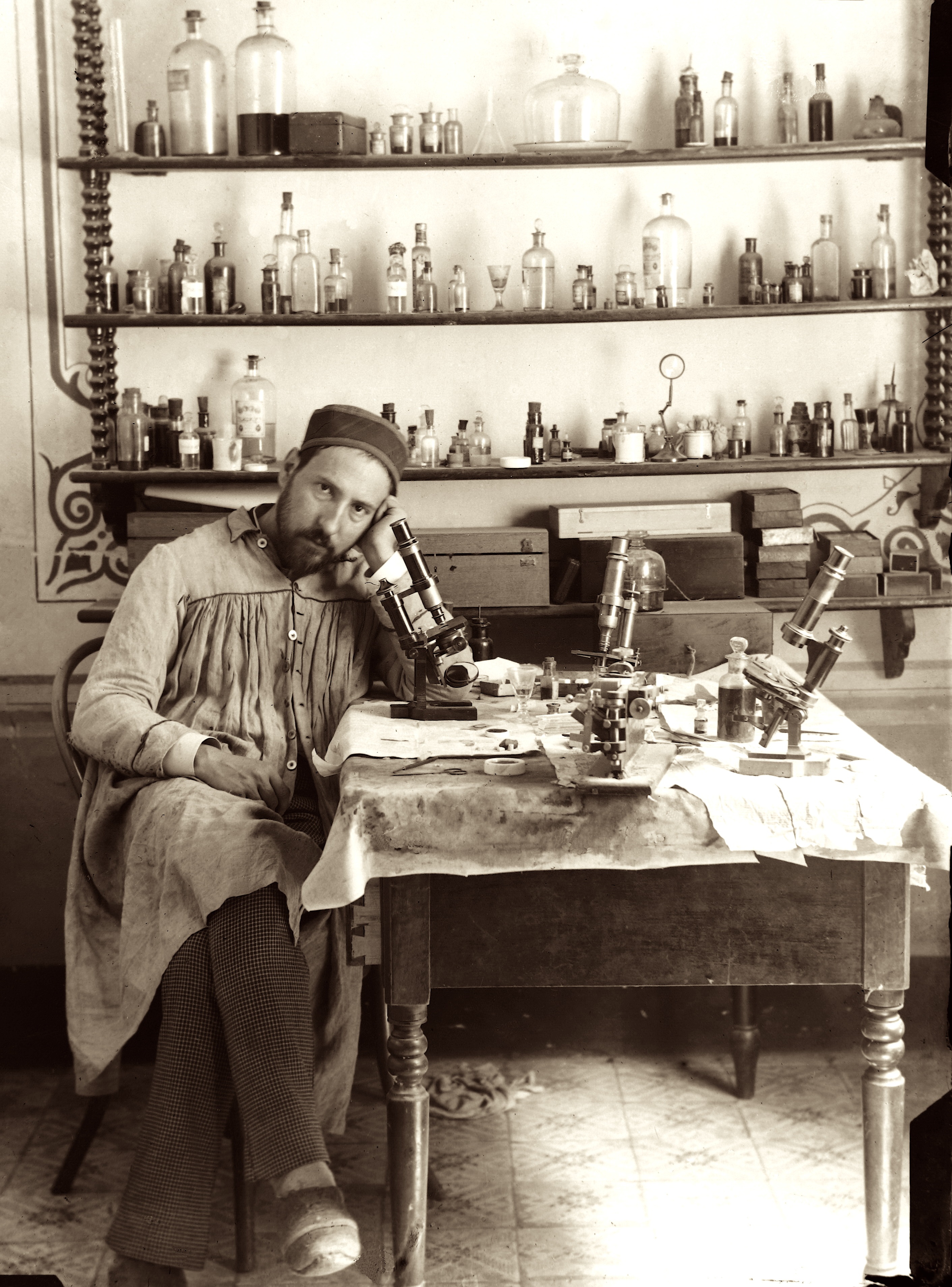
Santiago Ramón y Cajal
May 1, 1852 – October 17, 1934
| Div | |||||||||||||||
|---|---|---|---|---|---|---|---|---|---|---|---|---|---|---|---|
| |||||||||||||||
|
| Div | ||
|---|---|---|
| ||
| Div | ||||||||||
|---|---|---|---|---|---|---|---|---|---|---|
| ||||||||||
|

| Div | ||
|---|---|---|
| ||
Video
| Widget Connector | ||||||
|---|---|---|---|---|---|---|
|
| Div | ||
|---|---|---|
| ||
5th Installation (current)
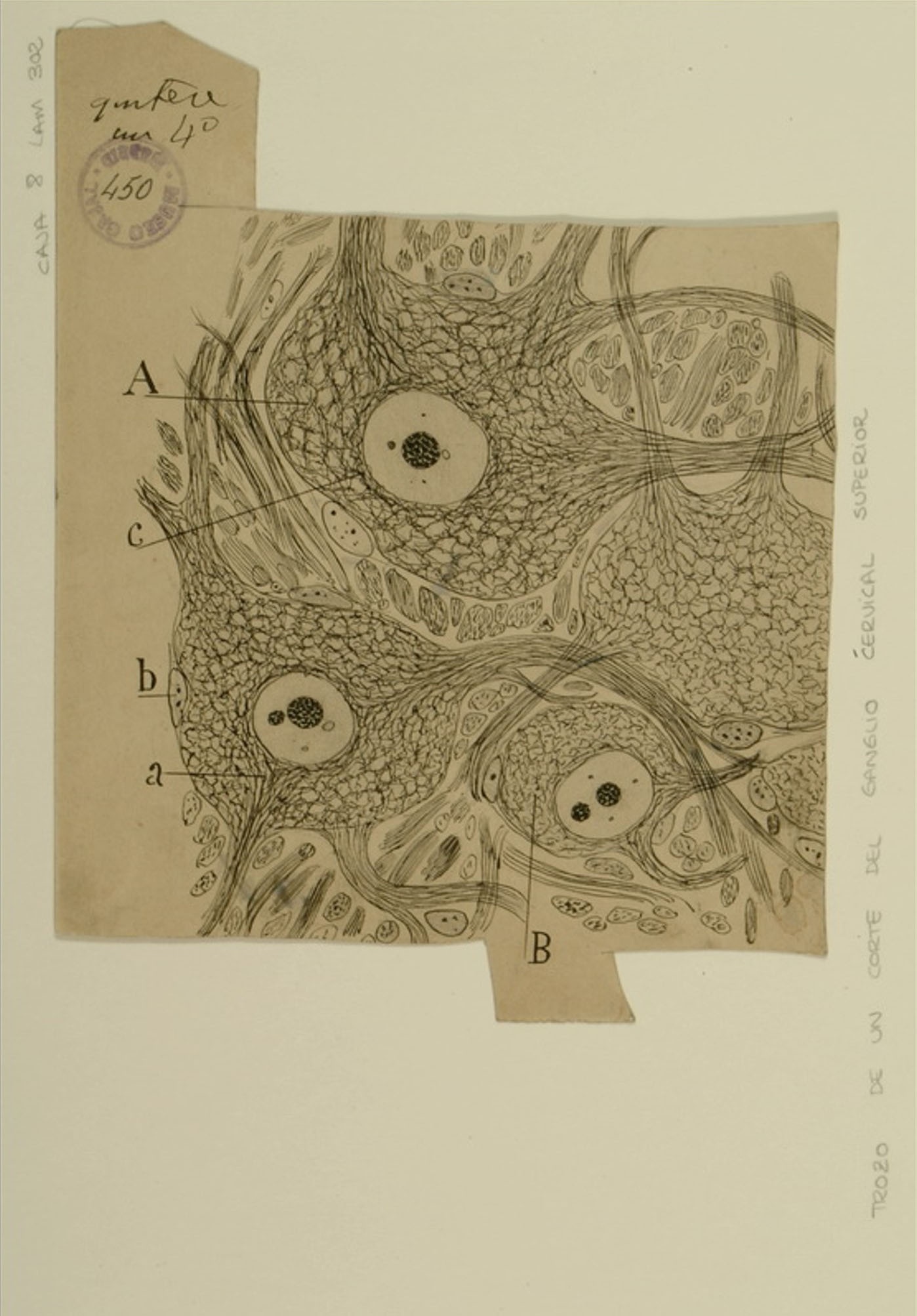
Upper cervical ganglia
| Div | ||||||||||||||||||||
|---|---|---|---|---|---|---|---|---|---|---|---|---|---|---|---|---|---|---|---|---|
| ||||||||||||||||||||
|
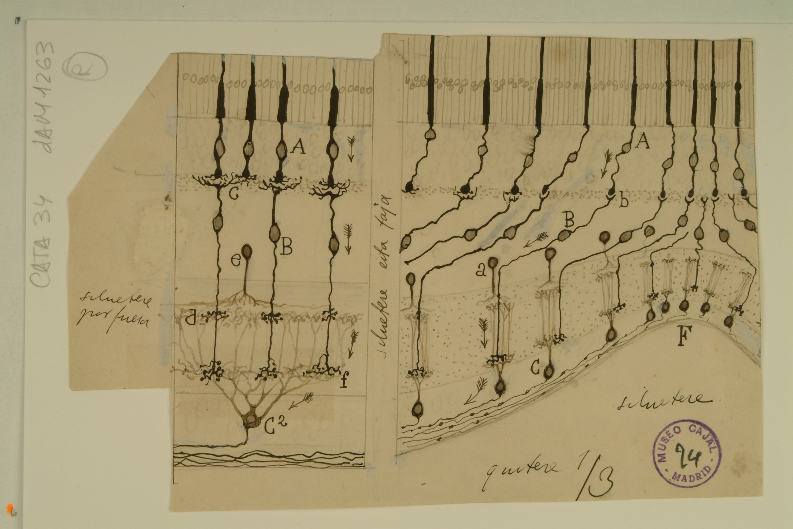
Comparison of visual and olfactory systems
| Div | ||||||||||||||||||||
|---|---|---|---|---|---|---|---|---|---|---|---|---|---|---|---|---|---|---|---|---|
| ||||||||||||||||||||
|

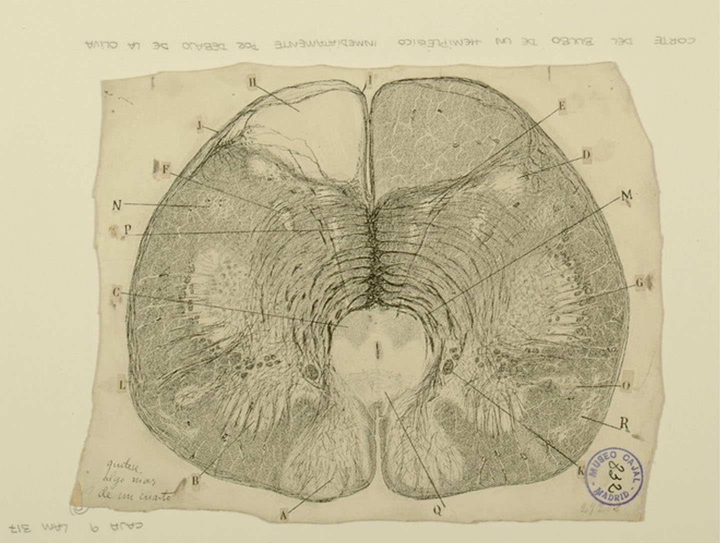
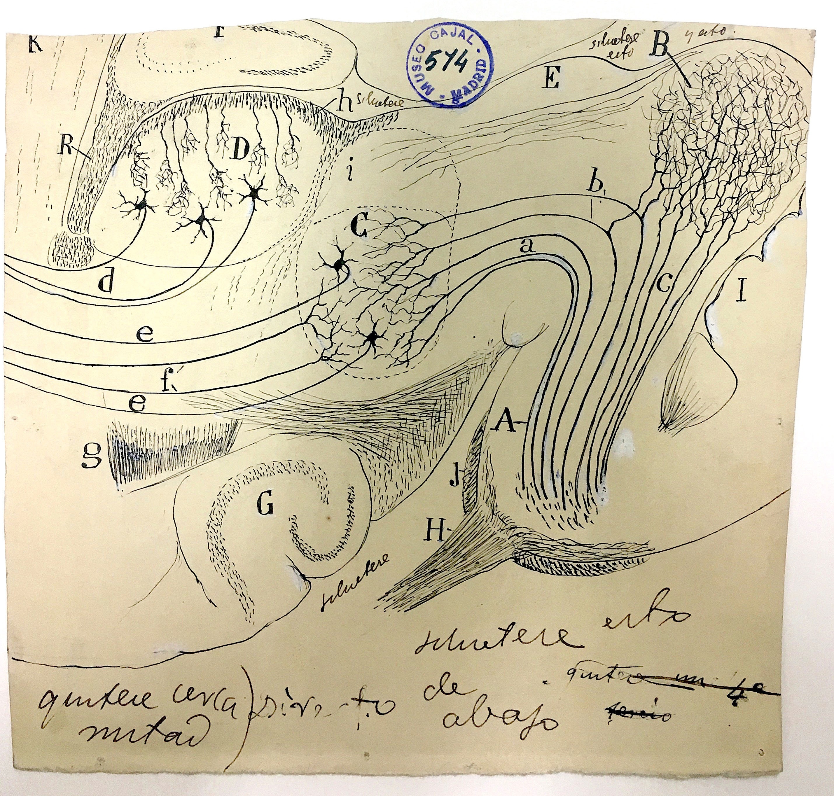
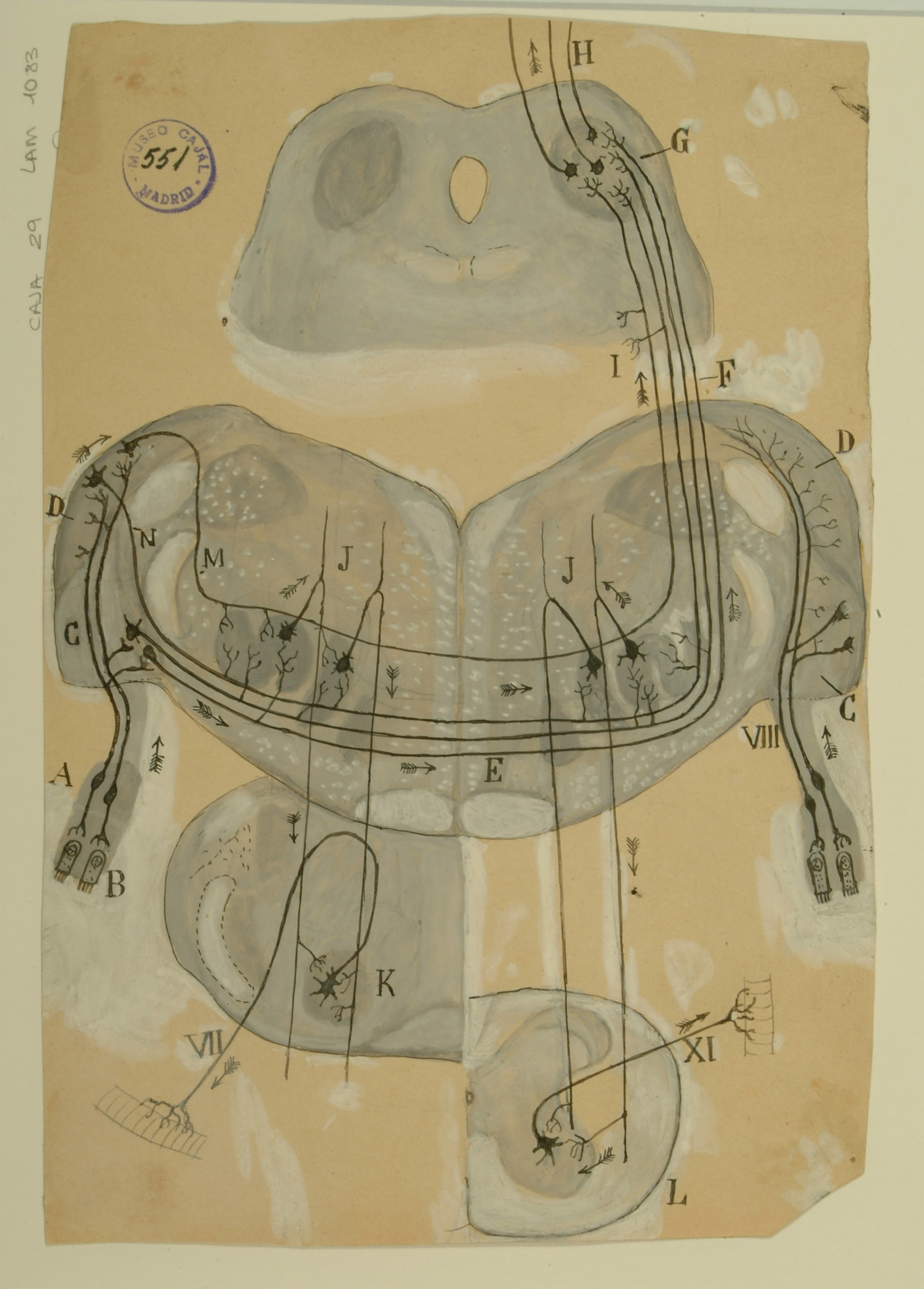
Medulla oblongata from a hemiplegic individual
| Div | ||||||||||||||||||||
|---|---|---|---|---|---|---|---|---|---|---|---|---|---|---|---|---|---|---|---|---|
| ||||||||||||||||||||
|
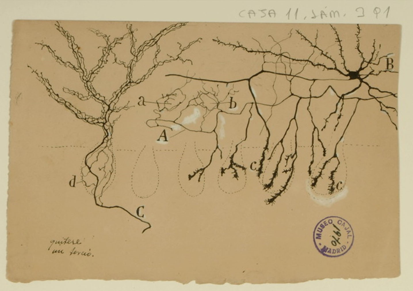
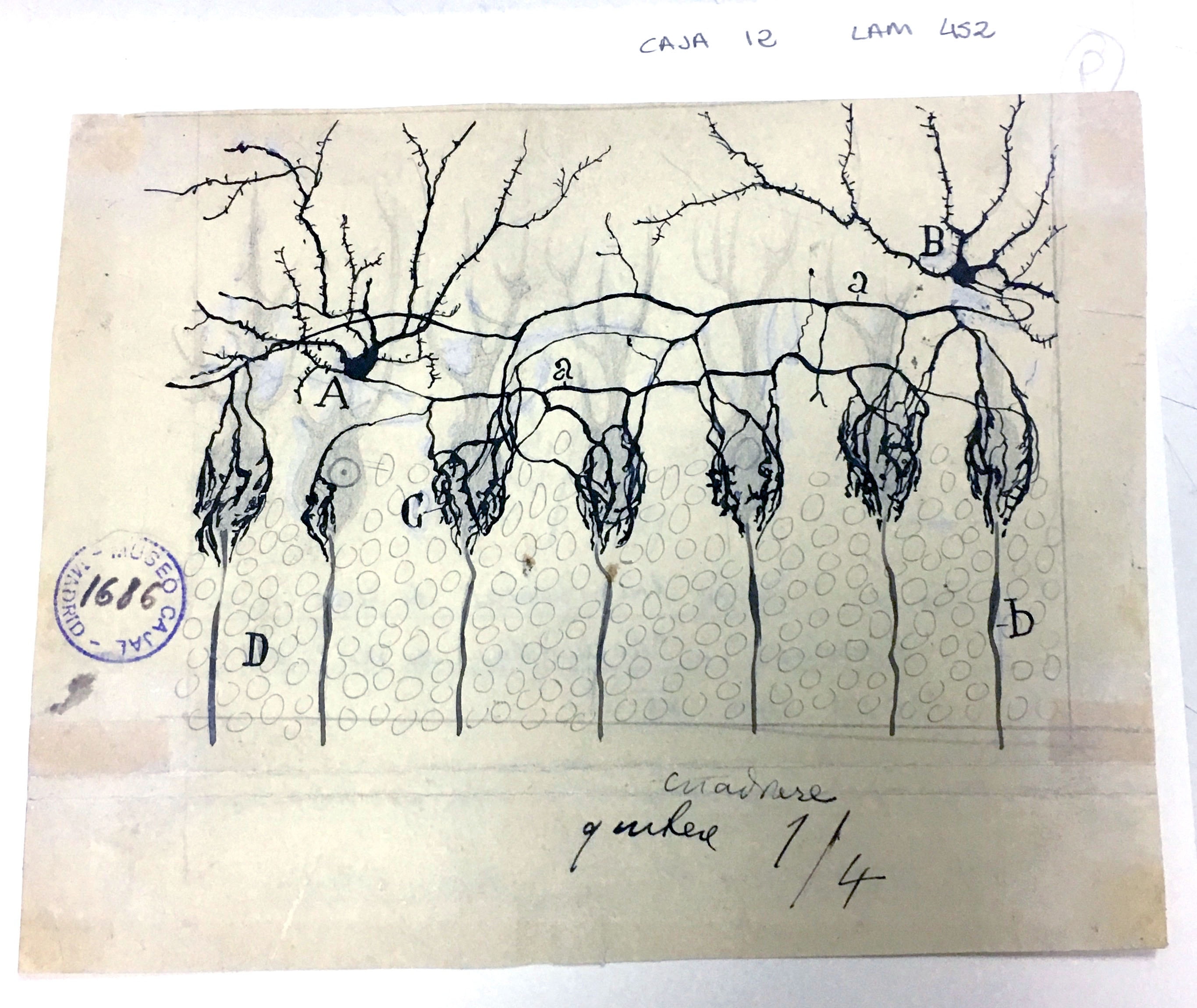
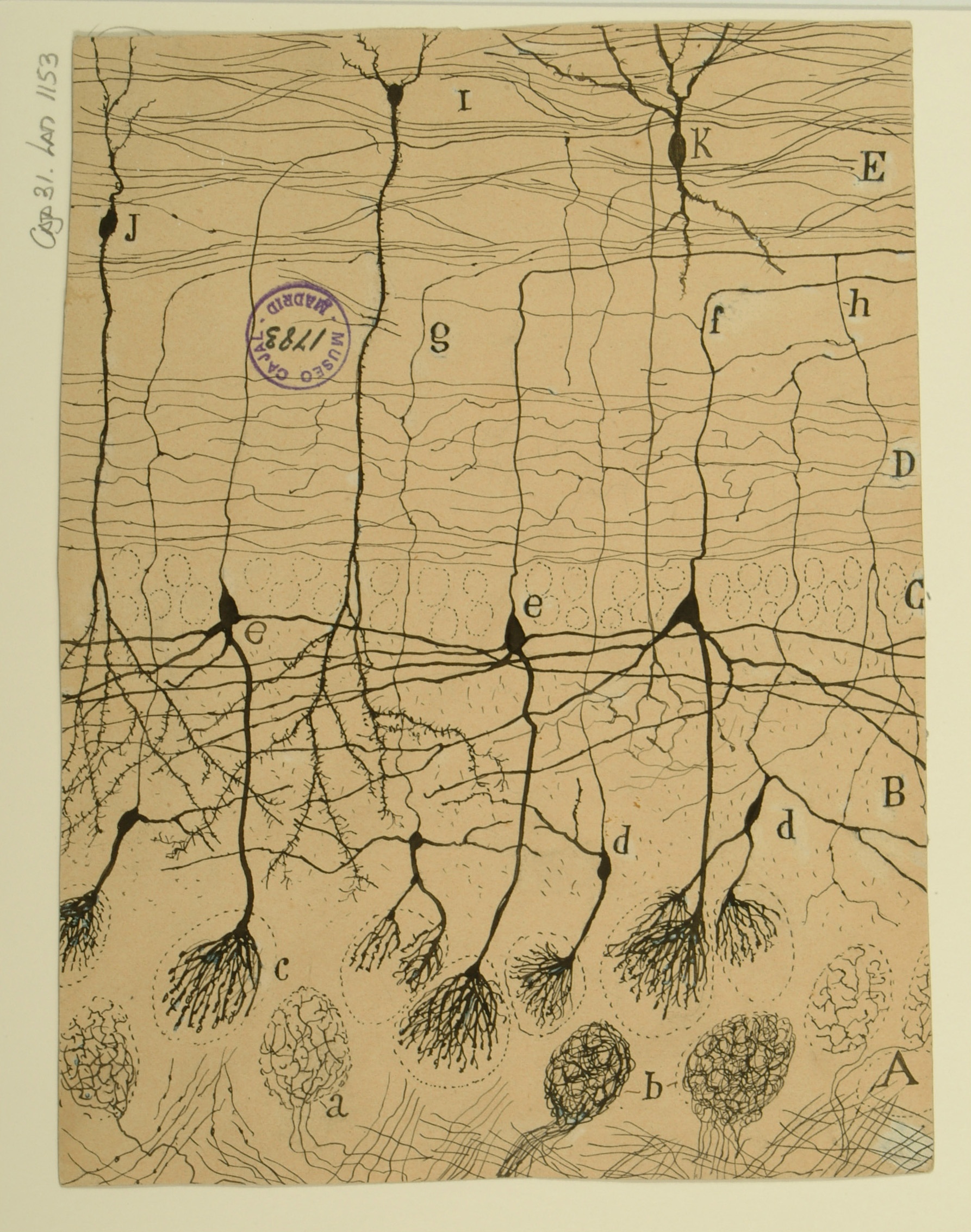
Innervation of Purkinje Cells
| Div | ||||||||||||||||||||
|---|---|---|---|---|---|---|---|---|---|---|---|---|---|---|---|---|---|---|---|---|
| ||||||||||||||||||||
|
Mouse olfactory bulb
| Div | ||||||||||||||||||||
|---|---|---|---|---|---|---|---|---|---|---|---|---|---|---|---|---|---|---|---|---|
| ||||||||||||||||||||
|
Mammalian retina
| Div | ||||||||||||||||||||
|---|---|---|---|---|---|---|---|---|---|---|---|---|---|---|---|---|---|---|---|---|
| ||||||||||||||||||||
|
Sphenoidal cortex in a 25-day old infant
| Div | ||||||||||||||||||||
|---|---|---|---|---|---|---|---|---|---|---|---|---|---|---|---|---|---|---|---|---|
| ||||||||||||||||||||
|

4th Installation
Nuclei in the auditory pathway
| Div | ||||||||||||||||||||
|---|---|---|---|---|---|---|---|---|---|---|---|---|---|---|---|---|---|---|---|---|
| ||||||||||||||||||||
|
| Div | ||
|---|---|---|
| ||
Coronal brain section of a young mouse
| Div | ||||||||||||||||||||
|---|---|---|---|---|---|---|---|---|---|---|---|---|---|---|---|---|---|---|---|---|
| ||||||||||||||||||||
|
| Div | ||
|---|---|---|
| ||
Basket cells in the cerebellum
| Div | ||||||||||||||||||||
|---|---|---|---|---|---|---|---|---|---|---|---|---|---|---|---|---|---|---|---|---|
| ||||||||||||||||||||
|
| Div | ||
|---|---|---|
| ||
Astrocytes at the border of a wound
| Div | ||||||||||||||||||||
|---|---|---|---|---|---|---|---|---|---|---|---|---|---|---|---|---|---|---|---|---|
| ||||||||||||||||||||
|

| Div | ||
|---|---|---|
| ||
Layer 5 of the visual cortex
| Div | ||||||||||||||||||||
|---|---|---|---|---|---|---|---|---|---|---|---|---|---|---|---|---|---|---|---|---|
| ||||||||||||||||||||
|

| Div | ||
|---|---|---|
| ||
Interneurons of the auditory cortex
| Div | ||||||||||||||||||||
|---|---|---|---|---|---|---|---|---|---|---|---|---|---|---|---|---|---|---|---|---|
| ||||||||||||||||||||
|

| Div | ||
|---|---|---|
| ||
Cells of the substantia gelatinosa of Rolando
| Div | ||||||||||||||||||||
|---|---|---|---|---|---|---|---|---|---|---|---|---|---|---|---|---|---|---|---|---|
| ||||||||||||||||||||
|

| Div | ||
|---|---|---|
| ||
3rd Installation
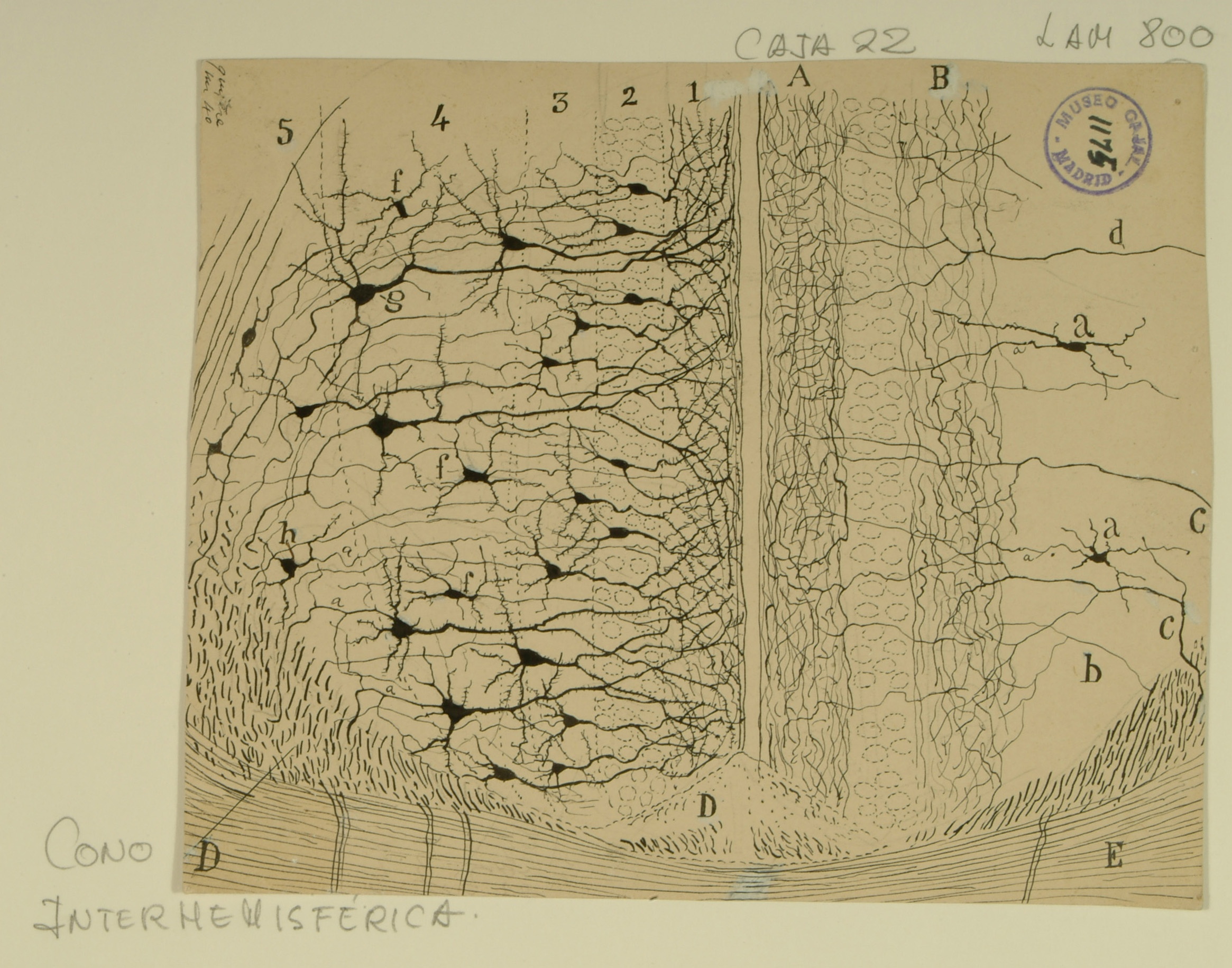
Cingulate Cortex
| Div | ||||||||||||||||||||
|---|---|---|---|---|---|---|---|---|---|---|---|---|---|---|---|---|---|---|---|---|
| ||||||||||||||||||||
|
| Div | ||
|---|---|---|
| ||
Diencephalic Nuclei
| Div | ||||||||||||||||||||
|---|---|---|---|---|---|---|---|---|---|---|---|---|---|---|---|---|---|---|---|---|
| ||||||||||||||||||||
|

| Div | ||
|---|---|---|
| ||
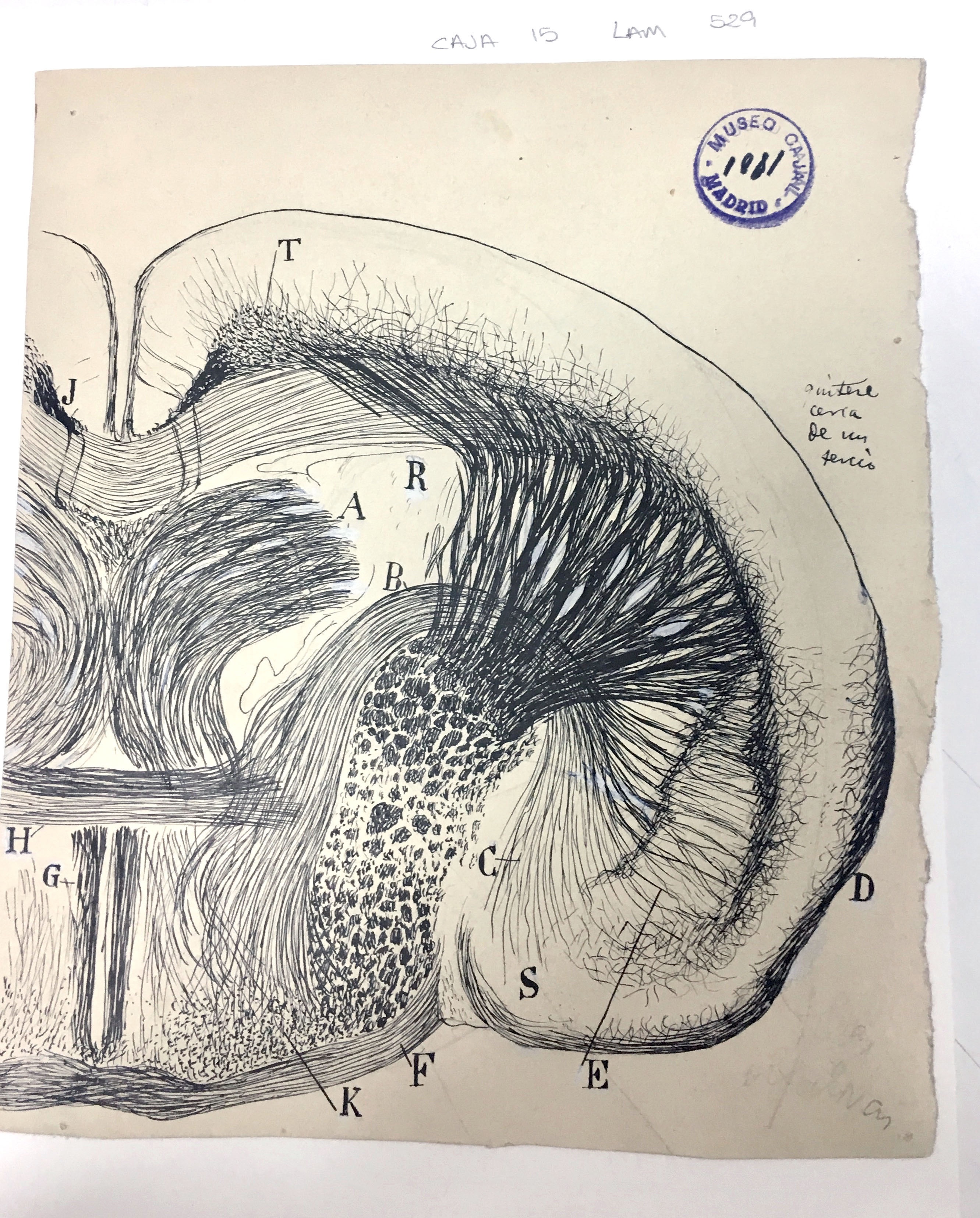
Trapezoid Body
| Div | ||||||||||||||||||||
|---|---|---|---|---|---|---|---|---|---|---|---|---|---|---|---|---|---|---|---|---|
| ||||||||||||||||||||
|
| Div | ||
|---|---|---|
| ||
Olfactory Bulb
| Div | ||||||||||||||||||||
|---|---|---|---|---|---|---|---|---|---|---|---|---|---|---|---|---|---|---|---|---|
| ||||||||||||||||||||
|
| Div | ||
|---|---|---|
| ||
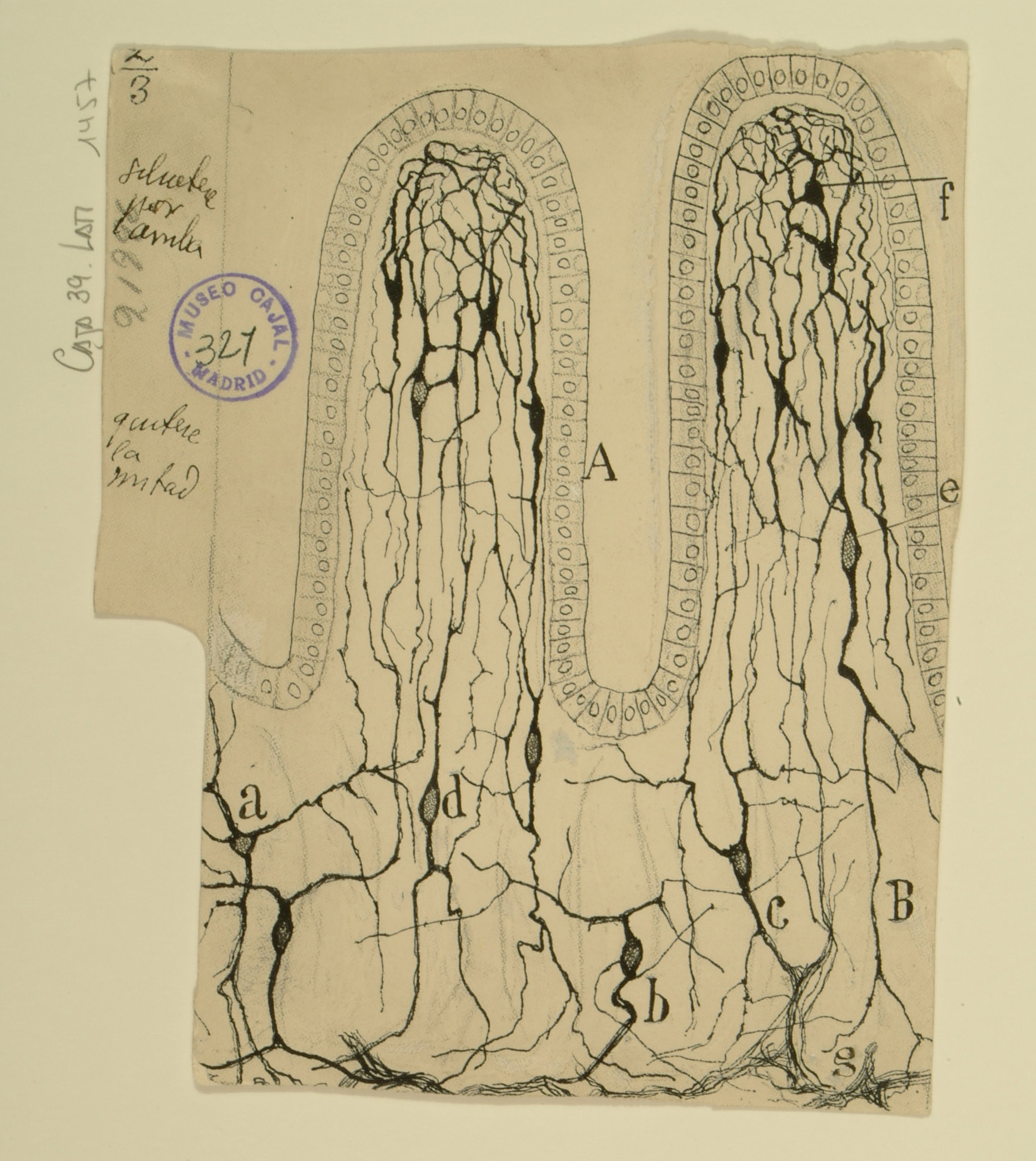
Intestinal Villi
| Div | ||||||||||||||||||||
|---|---|---|---|---|---|---|---|---|---|---|---|---|---|---|---|---|---|---|---|---|
| ||||||||||||||||||||
|
| Div | ||
|---|---|---|
| ||
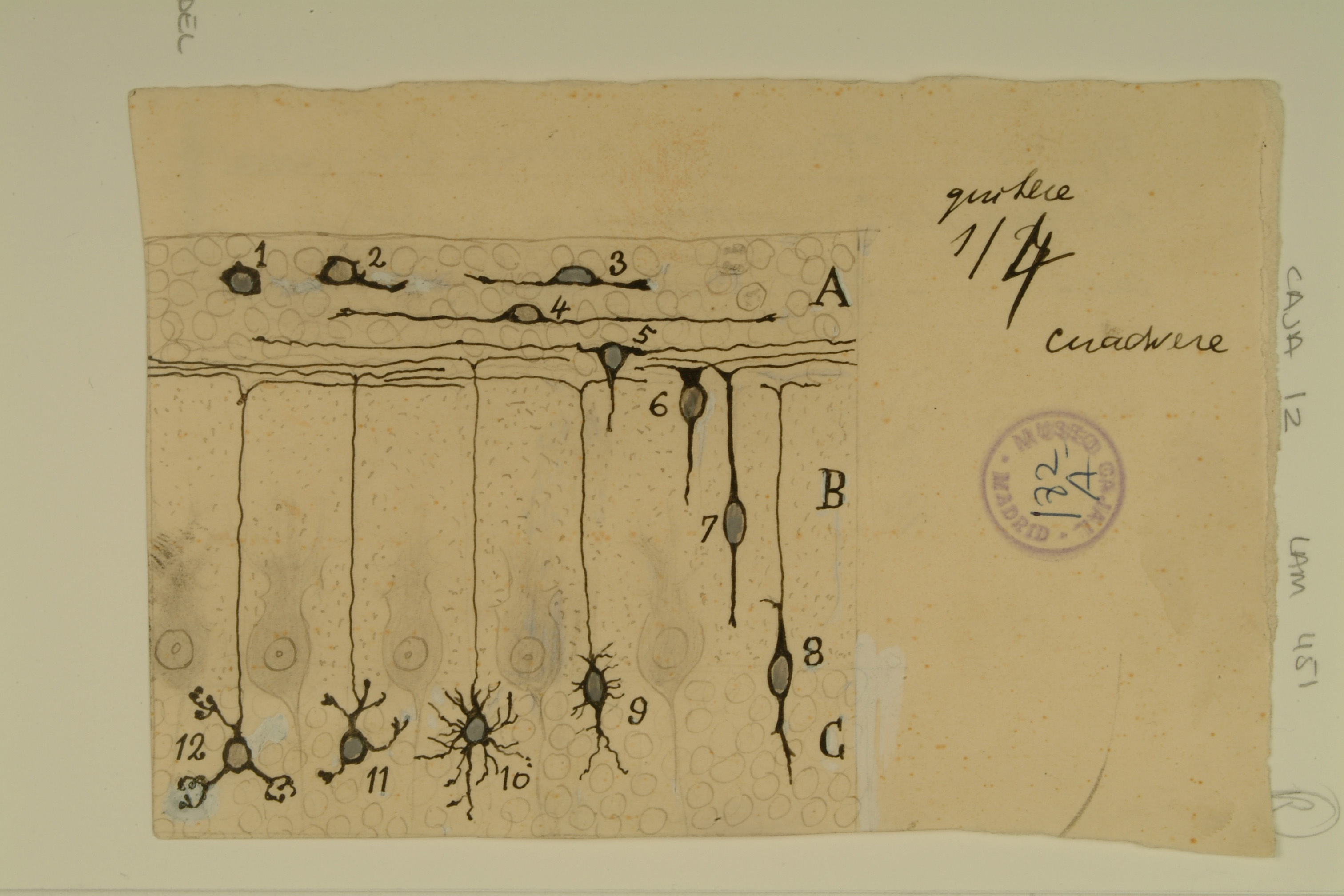
Development of Granule Cells in the Cerebellum
| Div | ||||||||||||||||||||
|---|---|---|---|---|---|---|---|---|---|---|---|---|---|---|---|---|---|---|---|---|
| ||||||||||||||||||||
|
| Div | ||
|---|---|---|
| ||
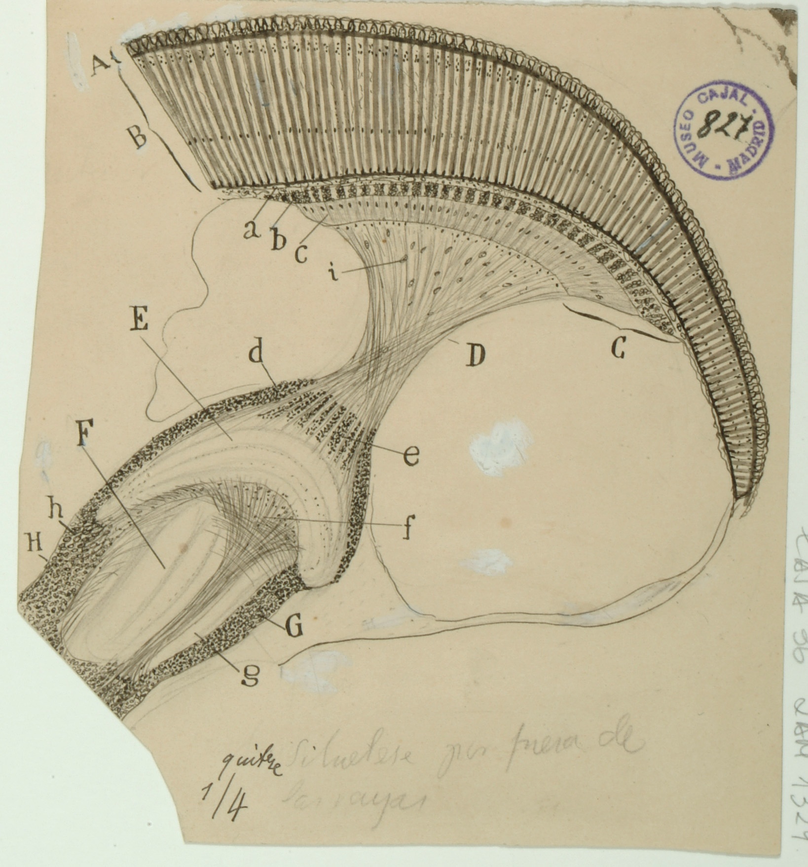
Fovea Centralis
| Div | ||||||||||||||||||||
|---|---|---|---|---|---|---|---|---|---|---|---|---|---|---|---|---|---|---|---|---|
| ||||||||||||||||||||
|
| Div | ||
|---|---|---|
| ||
2nd Installation
Growth Cones
| Div | ||||||||||||||||||||
|---|---|---|---|---|---|---|---|---|---|---|---|---|---|---|---|---|---|---|---|---|
| ||||||||||||||||||||
|

| Div | ||
|---|---|---|
| ||
Calyx of Held
| Div | ||||||||||||||||||||
|---|---|---|---|---|---|---|---|---|---|---|---|---|---|---|---|---|---|---|---|---|
| ||||||||||||||||||||
|

| Div | ||
|---|---|---|
| ||
Developing Neocortex
| Div | ||||||||||||||||||||
|---|---|---|---|---|---|---|---|---|---|---|---|---|---|---|---|---|---|---|---|---|
| ||||||||||||||||||||
|

| Div | ||
|---|---|---|
| ||
Olfactory System
| Div | ||||||||||||||||||||
|---|---|---|---|---|---|---|---|---|---|---|---|---|---|---|---|---|---|---|---|---|
| ||||||||||||||||||||
|

| Div | ||
|---|---|---|
| ||
Insect Visual System
| Div | ||||||||||||||||||||
|---|---|---|---|---|---|---|---|---|---|---|---|---|---|---|---|---|---|---|---|---|
| ||||||||||||||||||||
|
| Div | ||
|---|---|---|
| ||
Information Flow in the Retina
| Div | ||||||||||||||||||||
|---|---|---|---|---|---|---|---|---|---|---|---|---|---|---|---|---|---|---|---|---|
| ||||||||||||||||||||
|

| Div | ||
|---|---|---|
| ||
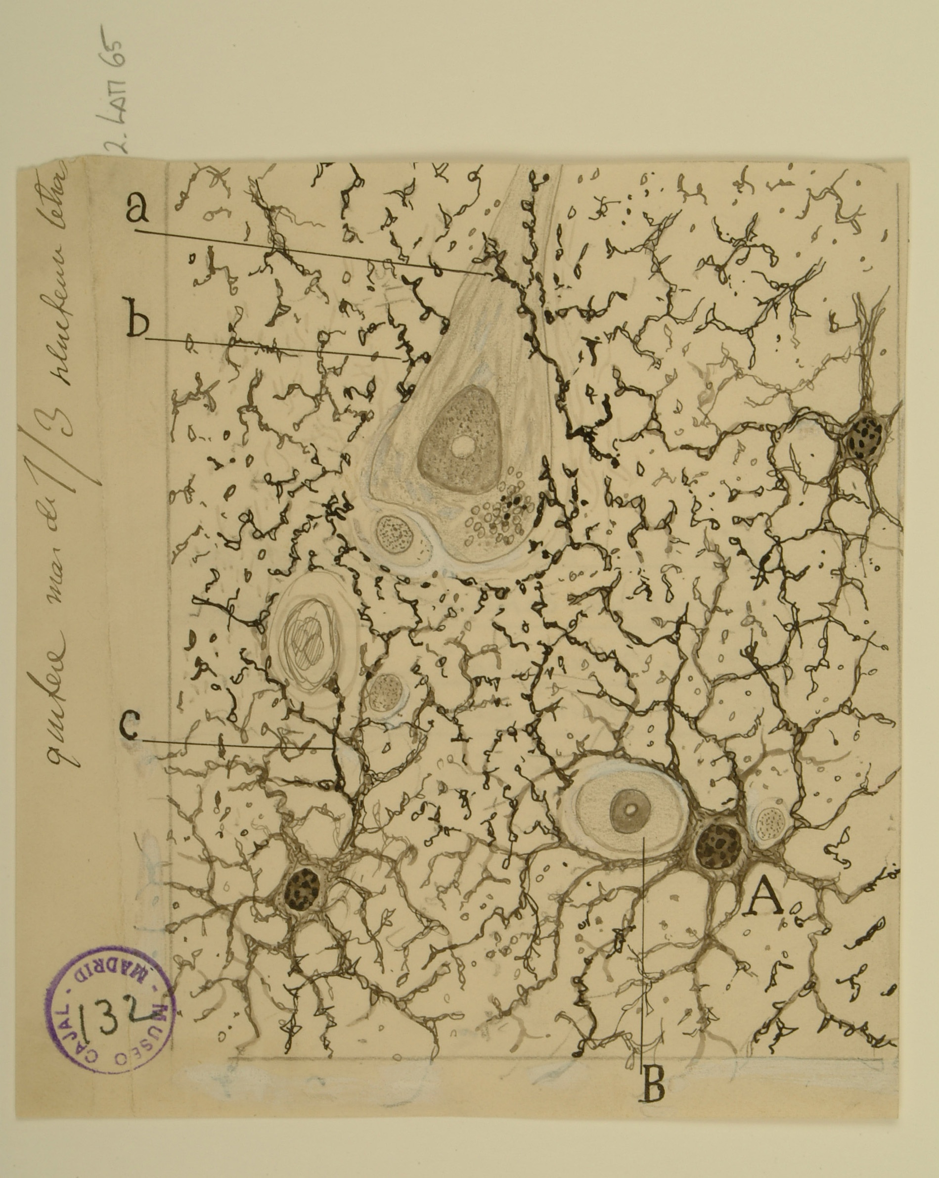
Astrocytes
| Div | ||||||||||||||||||||
|---|---|---|---|---|---|---|---|---|---|---|---|---|---|---|---|---|---|---|---|---|
| ||||||||||||||||||||
|
| Div | ||
|---|---|---|
| ||
1st Installation
Dentate Gyrus and CA3 Region of the Hippocampus
| Div | ||||||||||||||||||||
|---|---|---|---|---|---|---|---|---|---|---|---|---|---|---|---|---|---|---|---|---|
| ||||||||||||||||||||
|

| Div | ||
|---|---|---|
| ||
Hippocampus
| Div | ||||||||||||||||||||
|---|---|---|---|---|---|---|---|---|---|---|---|---|---|---|---|---|---|---|---|---|
| ||||||||||||||||||||
|

| Div | ||
|---|---|---|
| ||
Spinal Cord
| Div | ||||||||||||||||||||
|---|---|---|---|---|---|---|---|---|---|---|---|---|---|---|---|---|---|---|---|---|
| ||||||||||||||||||||
|

| Div | ||
|---|---|---|
| ||
Continuity vs. Contiguity
| Div | ||||||||||||||||||||
|---|---|---|---|---|---|---|---|---|---|---|---|---|---|---|---|---|---|---|---|---|
| ||||||||||||||||||||
|

| Div | ||
|---|---|---|
| ||
Cerebellum
| Div | ||||||||||||||||||||
|---|---|---|---|---|---|---|---|---|---|---|---|---|---|---|---|---|---|---|---|---|
| ||||||||||||||||||||
|

| Div | ||
|---|---|---|
| ||
Retina
| Div | ||||||||||||||||||||
|---|---|---|---|---|---|---|---|---|---|---|---|---|---|---|---|---|---|---|---|---|
| ||||||||||||||||||||
|

| Div | ||
|---|---|---|
| ||
Cortical Pyramidal Cells
| Div | ||||||||||||||||||||
|---|---|---|---|---|---|---|---|---|---|---|---|---|---|---|---|---|---|---|---|---|
| ||||||||||||||||||||
|

| Div | ||
|---|---|---|
| ||
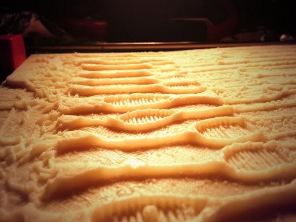
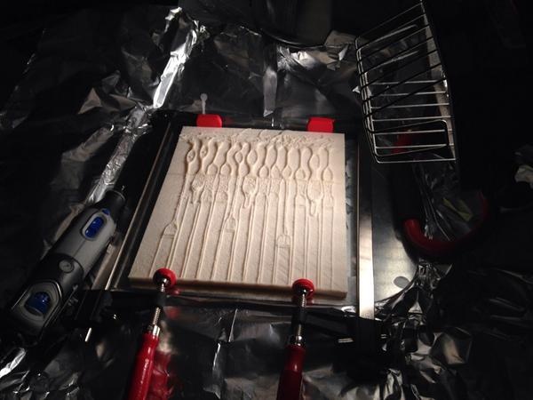
Test Print
This initial print file was created to create a process for making a 3d printable file from the image data. Two things became apparent in the creation of the file. One is that the relief, the distance between the background (generally paper without ink) and the foreground, neurons and structures inked on the paper, needed to be minimal in order to more effectively communicate the subject through touching with fingertips. If the relief was too high, as in this test print, then it became harder to feel small details.
| Span | ||
|---|---|---|
| ||
| Div | ||
|---|---|---|
| ||
Creating the 3D objects
Most of the 3D files were created by a process called "Height Displacement", in which the generally lighter background (absent of dark ink) represented the base, and the darker color would be raised "higher" from the background. Several other techniques were used in conjunction or instead of height displacement, including processing with software including Adobe Photoshop and Illustrator, 3D Coat, Blender, and Netfabb.
| Div | ||||||||||
|---|---|---|---|---|---|---|---|---|---|---|
| ||||||||||
|
Acknowledgements
This exhibition is a collaborative effort of the Office of NIH History’s Stetten Museum and the National Institute of Neurological Disorders and Stroke (NINDS). Jeffrey S. Diamond, Ph.D., Senior Investigator, Synaptic Physiology Section, Division of Intramural Research, NINDS, represented the NIH in negotiating the loan of the original drawings from the Cajal Institute of Madrid, Spain. NIH and the exhibition team are particularly grateful to Dr. Juan De Carlos, of the Cajal Institute for making this exhibition possible through the loan of the original drawings, and the archival images featured in the exhibition.
The exhibition team also thanks the National Library of Medicine, History of Medicine Division’s Paper Conservator, Holly Herro, for advising on appropriate exhibition light levels and mitigation strategies. Content and environmental design were created by Hank Grasso of the NIH Stetten Museum. Jamie Kugler, Ph.D., researched and composed the labels identifying for each of the four sets of (seven) drawings. This installation was produced under Chris Wanjek, OIR Director of Communications, Story Landis, Ph.D., (former) Director, NINDS, and Walter J. Koroshetz, M.D., (current Director, NINDS).
Overview
Content Tools
ThemeBuilder