Santiago Ramón y Cajal, a Spanish physician and scientist, was the first to describe the structure of the nervous system with exquisite precision. In what would become known as the “neuron doctrine,” he showed that the nervous system comprises individual cells (later termed “neurons”), that these cells connect to each other at small, specialized contact zones (now known as “synapses”), and that a single nerve cell typically possesses three anatomically distinct structures: the dendritic arbor, the cell body, and the axon. He further posited that neurons function as information processing units, using electrical impulses to communicate within functional networks. Cajal’s experimental work and theories provided the foundation for modern neurobiology.
An exhibition featuring revolving sets of seven original illustrations of famed scientist/artist Santiago Ramón y Cajal (on loan from the Instituto Cajal in Madrid, Spain), may be found near the North Entrance, on the first floor, of Building 35 on the NIH Campus.
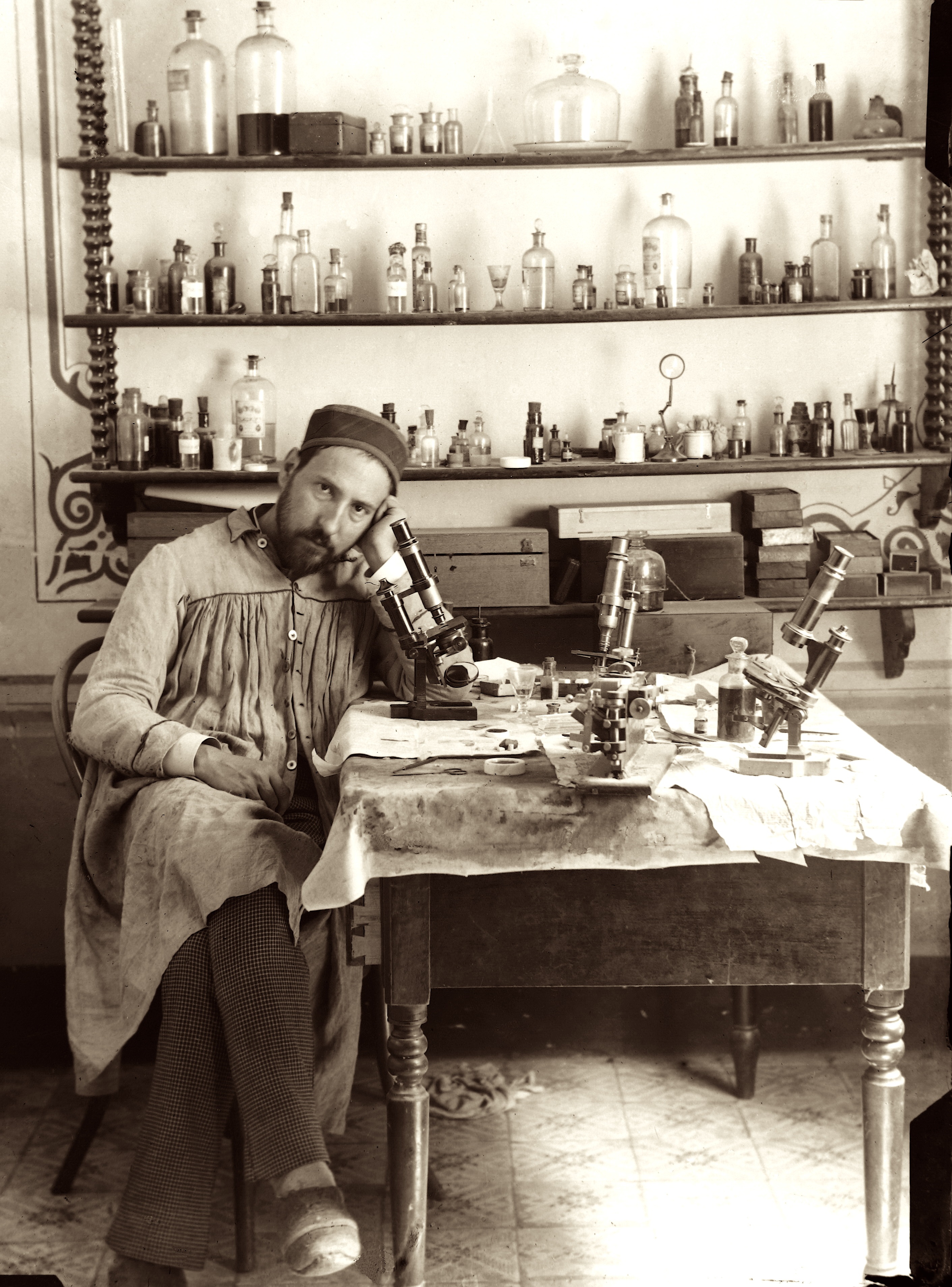
Cajal took this photograph of himself in his late-19thcentury laboratory (the shutter controller is cleverly hidden in his right hand). The array of chemicals and dyes he used to prepare tissue slides fill the shelves on the back wall. On his work table sit the microscopes through which he viewed cell structures, the art supplies that he used to render what he saw, and what appears to be a glass of sherry. In this single portrait, we see both the serious scientist and the studio artist. In 1906, Cajal and Camillo Golgi (the Italian physician-scientist who developed the tissue staining technique that Cajal used) shared the Nobel Prize in Physiology or Medicine “in recognition of their work on the structure of the nervous system.”
Div
idclass
hr-1
Div
class
hr-2
Div
class
hr-3
Div
class
usa-grid
Div
class
usa-width-one-half
Each featured original illustration from the early 1900s, is accompanied by a caption written to engage scientists, researchers and investigators who populate the NIH campus. As well as a 3-D printed rendering that enlarges a detail of the illustration above. In this way, the drawings are rendered more accessible to a variety of audiences—including vision-impaired visitors who can directly experience these tactile versions of Cajal's drawings. These files are made available on the 3D Print Exchange. Direct links to the 3-D print files are provided at the end of this page.
The Cajal illustrations currently on-view include: