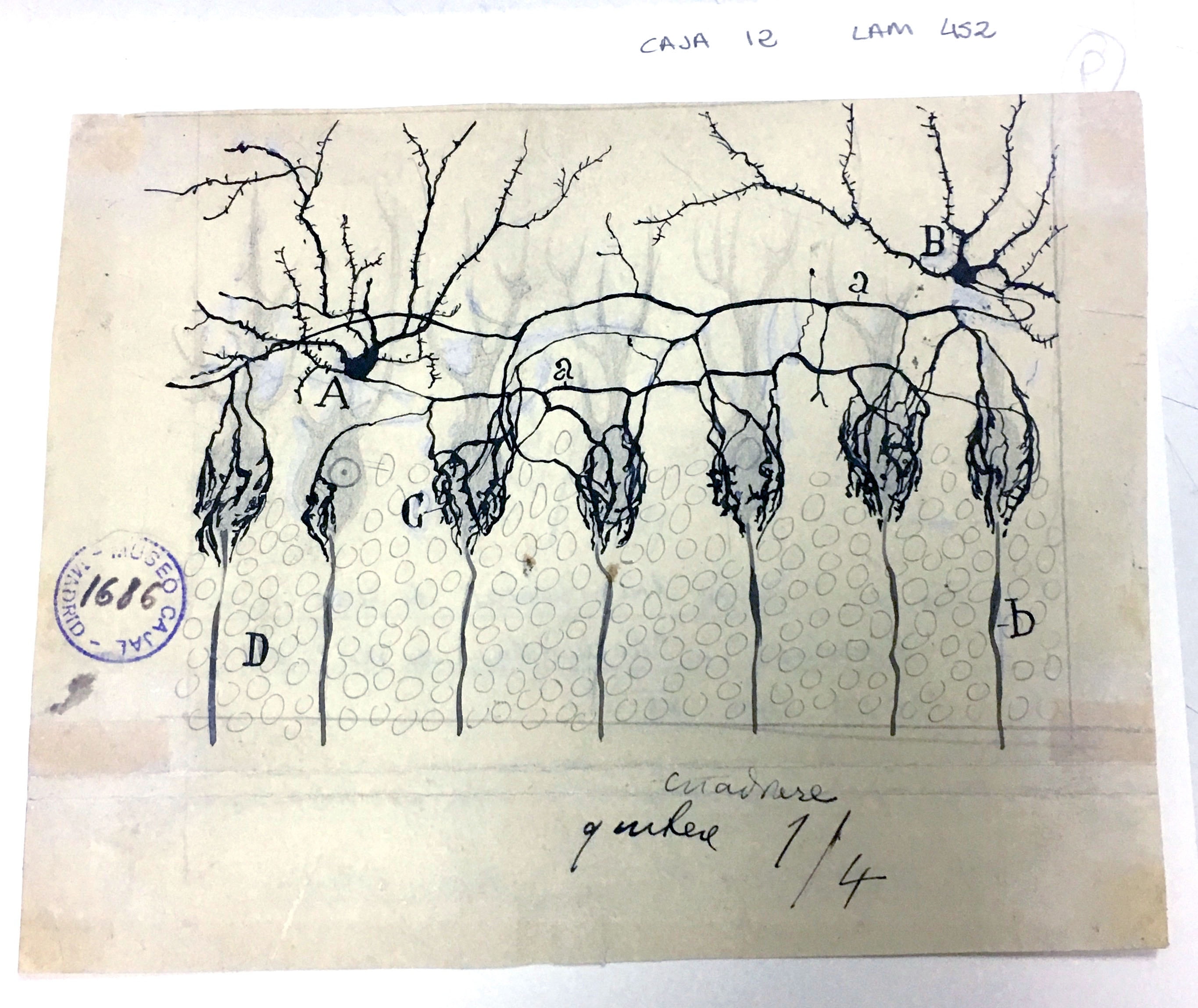
Basket cells are inhibitory interneurons found in several parts of the brain. Those shown here, in the cerebellum, make motor movement possible by preventing inhibitory signaling from Purkinje neurons. Each basket cell is composed of Purkinje neuron cell bodies surrounded by basketlike networks of axon branches (c) from the nearby stellate neurons (A and B); Cajal called these basketlike cell terminals ‘pinceau,’ French for ‘paintbrush.’
Using the silver nitrate staining method to visualize these cells, he recognized that although the axons of the stellate neurons made numerous synapses with the Purkinje neuron cell bodies, they did not fuse at any point. This supported his Neuron Doctrine, wherein the nervous system is composed of distinct cells rather than a network of continuously connected cells, and nervous impulses travel from the axon of one cell to the body of another.
Although he first posited the Neuron Doctrine in 1894, it was not until the 1950s, when the first electron microscopes became available, that scientists were able to confirm the existence of the synapse and thus validate Cajal’s theory.
Astrocytes are a type of macroglia that are critical for maintaining physiological homeostasis in the CNS and supporting neuronal function. Astrocytes in the grey and white matter of the brain typically have pedicles, or “feet”, that form contacts with capillaries (A, B, e) and control local blood flow.
Using a uranium-nitrate technique specifically for staining astrocytes on a tissue sample bordering a cerebral wound, Cajal observed not only normal astrocytes in contact with capillaries, but also small amoeboid cells (a,b,c). Other scientists, such as Alzheimer, had previously noted such cells in the CNS tissue of persons with various degenerative diseases, but their origins were uncertain. Cajal correctly inferred that these cells were astrocytes which had somehow reshaped themselves after the injury.
We now know that astrocytes become “reactive” after a brain injury: they become polarized, migrate, and their cell bodies swell. Such reactive astrocytes are postulated to have both beneficial (wound healing, limitation of inflammation) and detrimental (scar formation) roles in the response to injury.
Cajal inferred the flow of information between neurons from their structure and relative position. His ‘Law of Dynamic Polarization’ posits that each neuron is polarized: it has dendrites, through which signals are received, and an axon through which signals are transmitted to the dendrites of the next cells in the pathway. Thus, simply by observing the morphology and location of neurons in a tissue, he was able to discern the direction of signal transmission.
While this was relatively straightforward in the retina, which receives outside stimuli arrive from a particular direction and must be carried inward, Cajal’s real genius was revealed in the deduction of information flow in a tissue such as the hippocampus, where the sites of input and output were not immediately obvious.
The hippocampus, named for its resemblance to a seahorse (genus Hippocampus), comprises the Horns of Ammon (Cornu Ammonis, or CA regions), the dentate gyrus, and the subiculum. It receives signals from various parts of the brain via the entorhinal cortex, and signals flow through the hippocampus in the path shown by Cajal above (dentate gyrus CA3 gyrus → CA3 → CA1); its output travels through the fornix to the anterior thalamic nuclei and other destinations. Prior to Cajal’s observations, the fornix was thought to be a source of input to the hippocampus.