Versions Compared

Key

  • This line was added.
  • This line was removed.
  • Formatting was changed.

...

Div
classusa-grid
Div
classusa-width-one-half

Photograph of Santiago Ramón y CajalSantiago Ramón y Cajal

Span
classcredit

Courtesy of the Cajal Institute, Spanish National Research Council or CSIC©

Div
classusa-width-one-half

Santiago Ramón y Cajal, a Spanish physician and scientist, was the first to describe the structure of the nervous system with exquisite precision.  In what would become known as the “neuron doctrine,” he showed that the nervous system comprises individual cells (later termed “neurons”), that these cells connect to each other at small, specialized contact zones (now known as “synapses”), and that a single nerve cell typically possesses three anatomically distinct structures: the dendritic arbor, the cell body, and the axon. He further posited that neurons function as information processing units, using electrical impulses to communicate within functional networks.  Cajal’s experimental work and theories provided the foundation for modern neurobiology.

An exhibition featuring revolving sets of seven original illustrations of famed scientist/artist Santiago Ramón y Cajal (on loan from the Instituto Cajal in Madrid, Spain), may be found near the North Entrance, on the first floor, of Building 35 on the NIH Campus.

Cajal took this photograph of himself in his late-19thcentury laboratory (the shutter controller is cleverly hidden in his right hand). The array of chemicals and dyes he used to prepare tissue slides fill the shelves on the back wall. On his work table sit the microscopes through which he viewed cell structures, the art supplies that he used to render what he saw, and what appears to be a glass of sherry. In this single portrait, we see both the serious scientist and the studio artist. In 1906, Cajal and Camillo Golgi (the Italian physician-scientist who developed the tissue staining technique that Cajal used) shared the Nobel Prize in Physiology or Medicine “in recognition of their work on the structure of the nervous system.” 

...

Div
classusa-grid
Div
classusa-width-one-half

Hand drawn illustration of Nuclei in the auditory pathway

Span
classcredit

Courtesy of the Cajal Institute, Spanish National Research Council or CSIC©

Div
classusa-width-one-half
The mammalian auditory midbrain, shown above, is part of the ascending auditory pathway, responsible for relaying sensory signals from the ear into the primary auditory cortex deep in the brain.  Cajal’s microscopy studies led him to believe that the lateral leminiscus (A) received input from the cochlear and superior olivary nuclei, and carried some of it to the inferior colliculus (B), which integrated the signals necessary for auditory reflexes, while the bulk of the information was sent directly to the medial geniculate body (C), which then relayed the information on to the auditory cortex via the thalamo-cortical path (e).  

Modern studies have shown, however, that the inferior colliculus actually processes nearly all the input sent to the medial geniculate body and receives signals from the descending auditory pathway, as well as providing the motor integration necessary for auditory reflexes hypothesized by Cajal, making it a true hub for auditory signaling.

...

Div
classusa-grid
Div
classusa-width-one-half

Span
classcredit

Courtesy of the Cajal Institute, Spanish National Research Council or CSIC©

Div
classusa-width-one-half

The lenticular nucleus (E) is a lens-shaped bundle of neurons that, along with the caudate nucleus (R) and the internal capsule, comprises the corpus striatum.  Cajal used this drawing in his Texture of the Nervous System of Man and Vertebrates to illustrate the relatively large size of the lenticular nucleus in small mammals – in this case, a mouse – as compared to humans.  

Although Cajal posited that the corpus striatum in general was of decreasing evolutionary importance and only useful for the coordination of higher reflexes, we now know that it is important for the facilitation of voluntary movement.  The complexity and attention to detail in this drawing showcase Cajal’s skill in translating the view through his microscope lens to the page, where the structures he depicts are easily identifiable to today’s scientists more than 100 years after he put ink to paper.


Span
classusa-button

3dpx-010424 - Axonal Tracts

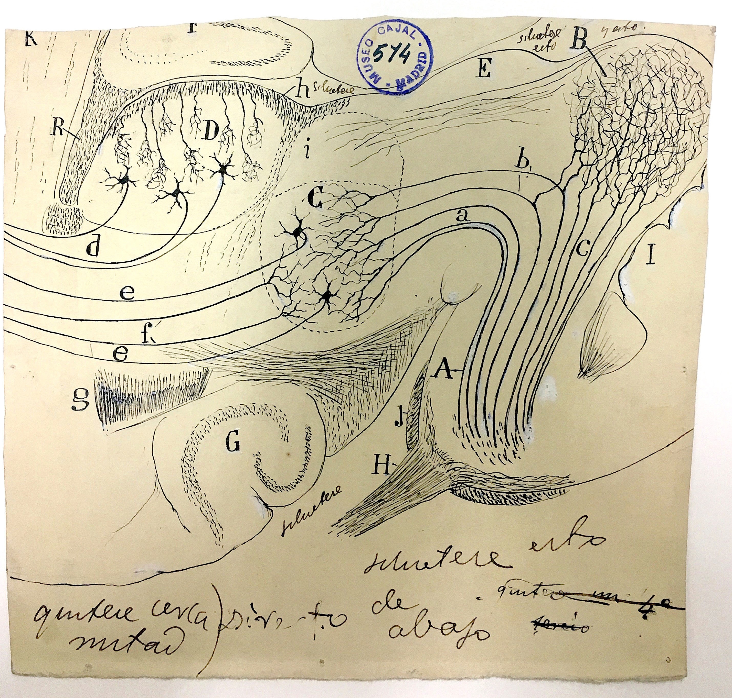
Basket cells in the cerebellum

...

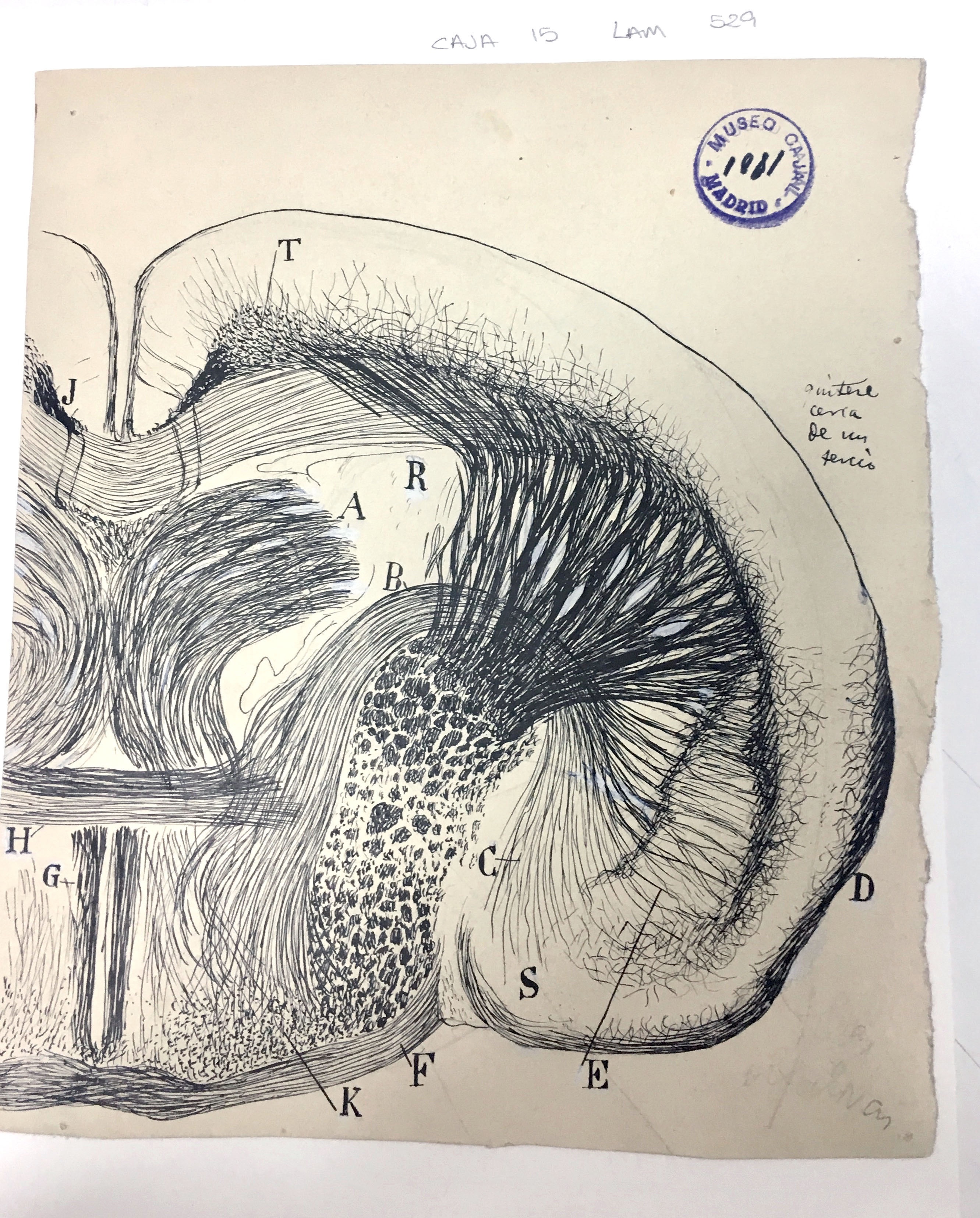
This initial print file was created to create a process for making a 3d printable file from the image data. Two things became apparent in the creation of the file.  One is that the relief, the distance between the background (generally paper without ink) and the foreground, neurons and structures inked on the paper, needed to be minimal in order to more effectively communicate the subject through touching with fingertips. If the relief was too high, as in this test print, then it became harder to feel small details.


Span
classusa-button

3DPX-000789 - Cortical Pyramidal Cells

...

The exhibition team also thanks the National Library of Medicine, History of Medicine Division’s Paper Conservator, Holly Herro, for advising on appropriate exhibition light levels and mitigation strategies.  Content and environmental design were created by Hank Grasso of the NIH Stetten Museum.  Jamie Kugler, Ph.D., researched and composed the labels identifying for each of the four sets of (seven) drawings.  This installation was produced under Chris Wanjek, OIR Director of Communications, Story Landis, Ph.D., (former) Director, NINDS, and Walter J. Koroshetz, M.D., (current Director, NINDS).

...

classusa-grid

...