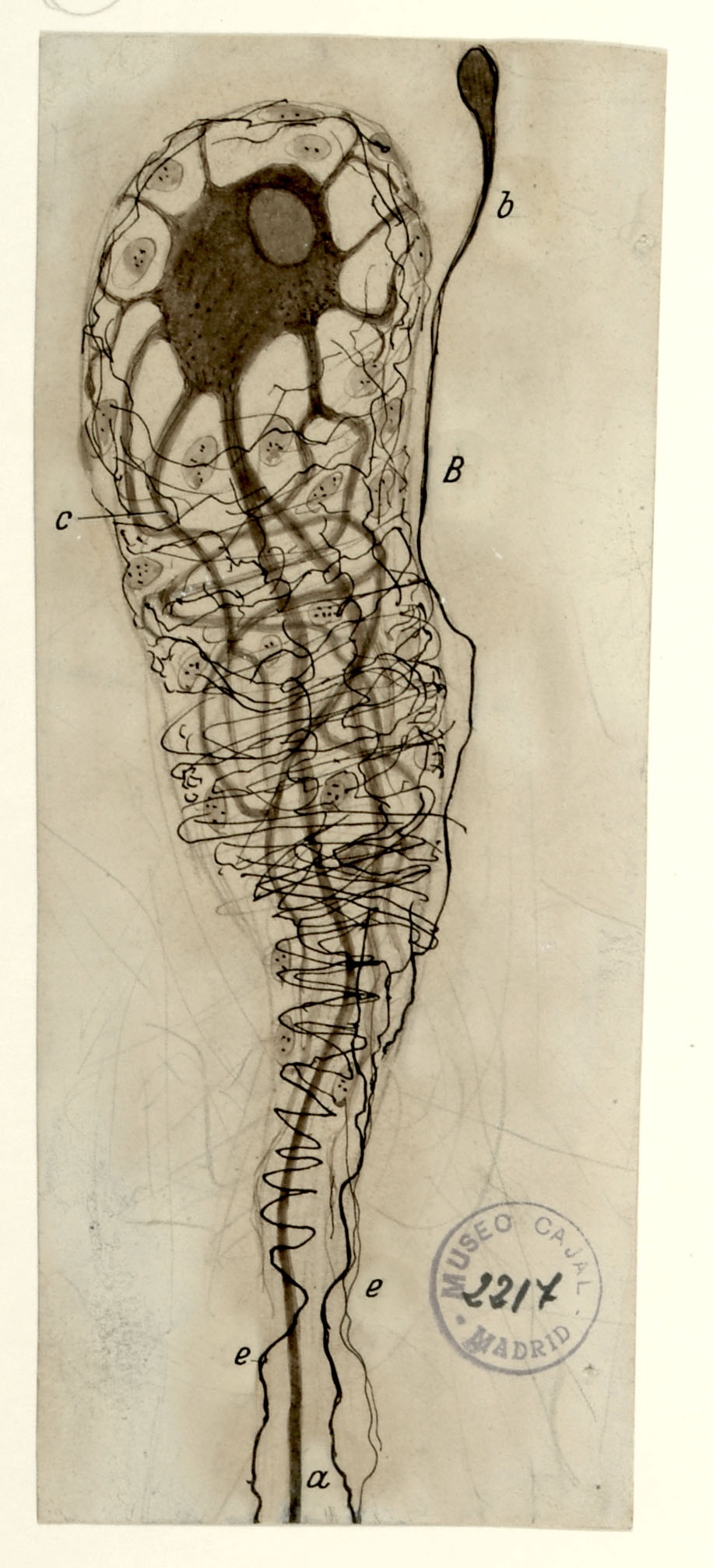
In 1905, Cajal studied human sympathetic ganglia and found morphological arrangements he had not yet seen in other species. Sympathetic ganglia comprises the thousands of afferent and efferent nerve cell bodies that run along either side of the spinal cord, connecting major organ systems, such as the renal system, to the spinal cord and brain. The kidney, a main organ within the renal system, filters blood to remove toxins via millions of structures called glomeruli, consisting of a tuft of blood vessels surrounded by a cuplike cellular structure known as Bowman’s Capsule. Shown above is a single neuron innervating a single glomerulus from a 50-year-old human subject, with a distinctive “comet” shape comprised of a very rich periglomerular nerve arborization. The kidney is innervated by both sympathetic and parasympathetic fibers; the innervating parasympathetic fibers originate from the vagal nerve. Pain signals caused by kidney stones may activate cross signaling along the vagal route, causing the well-known nausea and vomiting associated with kidney stones via the pathway shown in drawing no. 5.
During cerebellar development, climbing fibers reach the Purkinje cell bodies, making perisomatic contacts. Later they reach the dendrites and the contacts they make with these cells are perisomatic and peridendritic and, at the end of their development, the contacts are reduced to exclusively peridendritic.
In 1890, Cajal published a paper (Sobre la existencia de células nerviosas especiales en la primera capa de las circunvoluciones cerebrales. Gaceta médica catalana, nº 23: 737-739) reporting the existence of special nerve cells in the first layer of the cerebral cortex of lower mammals. He called them special because they had a unique peculiarity: they had two or more axons. Soon after, Gustav Retzius described the same cells in higher mammals and primates. Since then, they have been known as Cajal-Retzius cells. Today it is known that they are the first cell population to appear in the developing cerebral cortex and their importance in the stratification of this structure, through the secretion of the reelin protein.
In 1891, Cajal enunciated his Law of Dynamic Polarization at a Medical Congress in the city of Valencia. This Law says that the nervous impulse is polarized, going in only one direction. This is so, he explained, because the neuron receives the nerve impulse in its dendrites, transports it to the cell body and releases it through the terminal buttons of its axon, which come into contact with the dendrites of other cells. However, studying the optic tectum of birds in 1892, he found neurons whose axon does not emerge from the cell body, but from one of its dendrites, so his theory of Dynamic Polarization could not be fulfilled. He studies this phenomenon and realizes that, in these cases, the nerve impulse did not need to reach the cell body and have to go back through the dendrite to be able to exit through the axon, but was released directly through the axon without having to first reach the cell body. He then formulates a variation to his theory of Dynamic Polarization, which he enunciates as Theory of Axipetal Polarization.
This diagram details the body-spanning circuit underlying cough and vomiting. Irritation of sensory neurons in the larynx (A) or the stomach (J) initiates signals that travel through the vagus nerve (B) to pre-motor neurons (M, D), and onwards to motor neurons (F,H) that then control the movement of the chest and abdominal wall muscles (K) or the smooth muscle of the stomach (I), respectively. The cough reflex is extremely important for clearing the lungs of impediments that inhibit their gas-exchanging function. Its dysregulation can lead to damage of the airway mucosa and degrade quality of life. New targets for cough-suppressant drugs are being sought in the sensory neurons in the lungs and throat (A) and include blockers of transmembrane channels aimed at reducing nerve excitability.
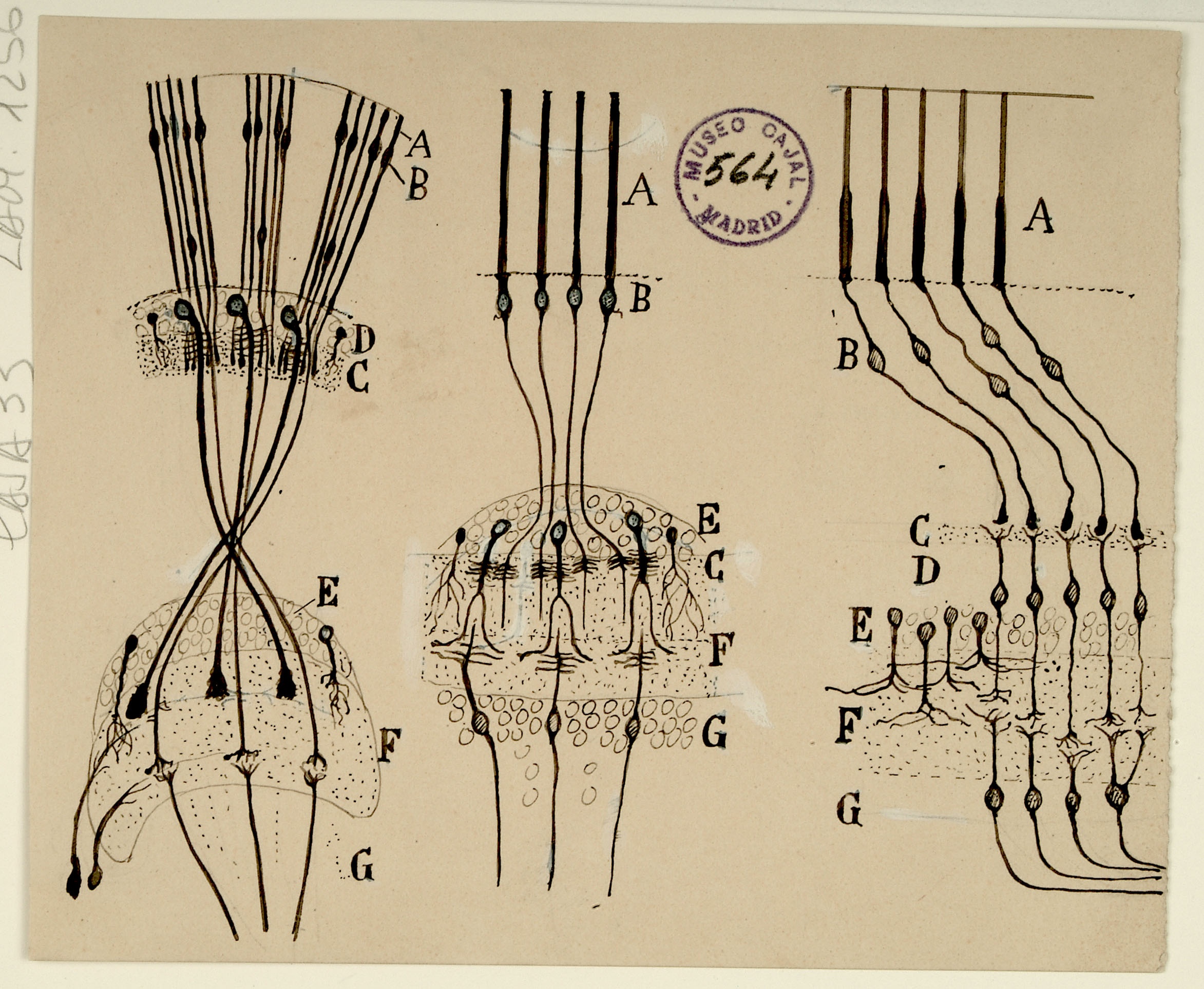
Cajal’s fascination with the retina is well documented – his publications on retinal neurons and organization in various species span 45 years, the length of his career. Shown above are schematics of insect (left), cephalopod (middle), and vertebrate (right) retinas, highlighting their similarities. All of these retinas are composed of photoreceptors (A) which are activated by light and transmit signals through synapses of intermediary cell types (B, C, F) onwards toward deeper brain processing (G).
Cajal has oriented all these diagrams with the photoreceptors at the top for ease of comparison, but in fact the diagram of the vertebrate retina is more correctly shown inverted compared to the other two, with its photoreceptors located at the bottom of the diagram. This is because the photoreceptors of invertebrates, such as insects and cephalopods, are exposed to light directly as the top layer of the retina, while the photoreceptors of vertebrates are actually at the bottom of the retina. Light must pass through the transparent layers of nerves above them before reaching them.
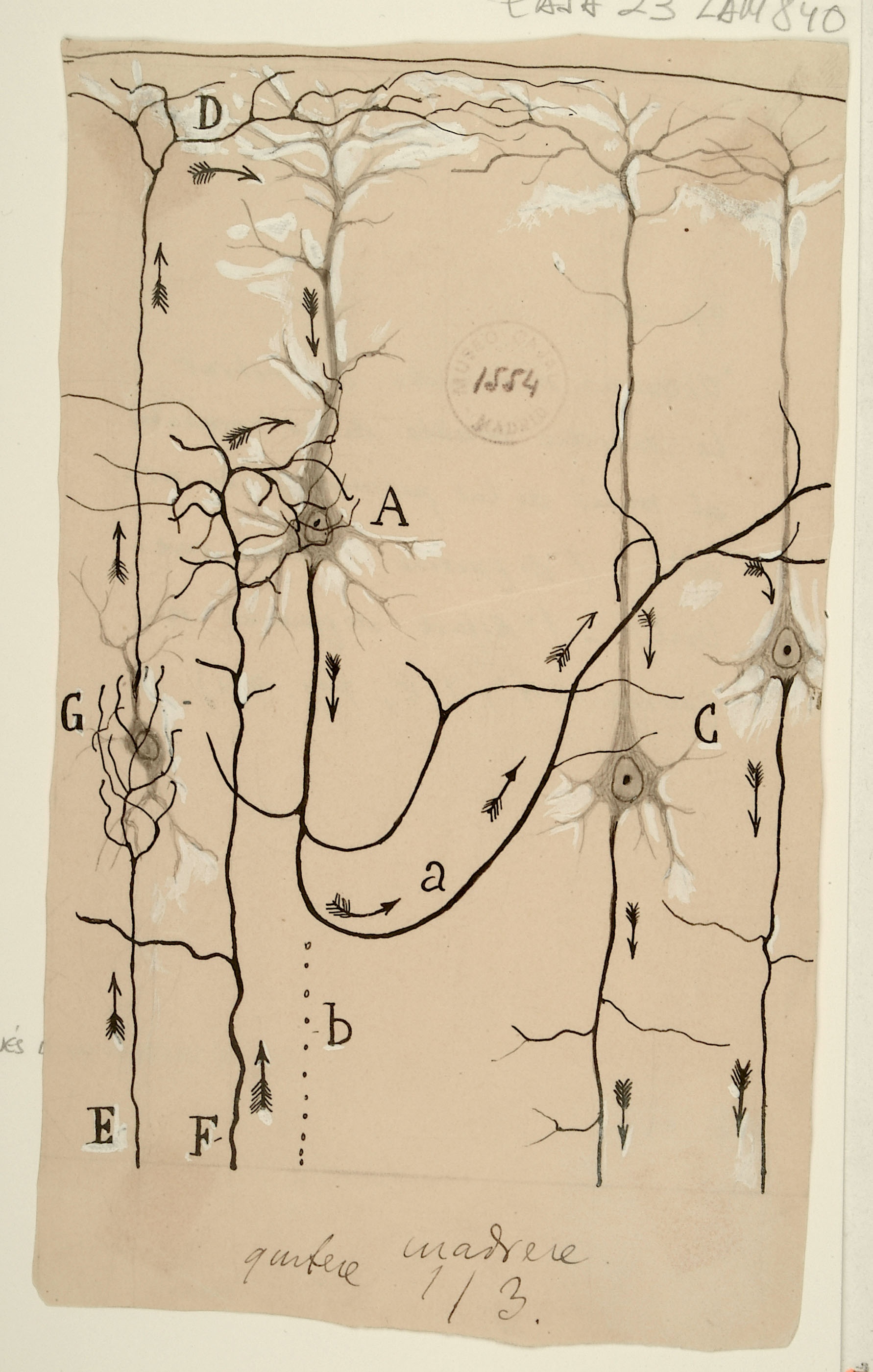
Here, Cajal illustrates a possible flow of information within the cerebral cortex, focusing on 4 pyramidal neurons. Inputs (E,F) come in several forms, including projections to apical dendrites (D) and to intermediate layers (A). After relay through other cortical neurons (a), deeper layer neurons (C) project out of the cortex. While there is strong anatomical evidence for these canonical paths of information flow, in recent years we have learned that the high degree of connectivity and diversity of cells in the cortex makes the fundamental computation of the cortex quite complex: a single cortical column has hundreds of thousands of such pyramidal cells, connected by millions of synapses. Cajal’s depiction of this circuit presages the 'canonical cortical microcircuit' of Douglas and Martin by nearly 100 years. The field is currently engaged in an effort to extend the canonical circuit ideas to the entire cortical network: understanding how the massive local connectivity builds on this canonical circuit backbone to create cortical computation.