Versions Compared

Key

  • This line was added.
  • This line was removed.
  • Formatting was changed.

...

Div
classgrid-row grid-gap
Div
classdesktop:grid-col-6

In 1891, Cajal enunciated his Law of Dynamic Polarization at a Medical Congress in the city of Valencia. This Law says that the nervous impulse is polarized, going in only one direction. This is so, he explained, because the neuron receives the nerve impulse in its dendrites, transports it to the cell body and releases it through the terminal buttons of its axon, which come into contact with the dendrites of other cells. However, studying the optic tectum of birds in 1892, he found neurons whose axon does not emerge from the cell body, but from one of its dendrites, so his theory of Dynamic Polarization could not be fulfilled. He studies this phenomenon and realizes that, in these cases, the nerve impulse did not need to reach the cell body and have to go back through the dendrite to be able to exit through the axon, but was released directly through the axon without having to first reach the cell body. He then formulates a variation to his theory of Dynamic Polarization, which he enunciates as Theory of Axipetal Polarization.

Div
classdesktop:grid-col-6

Span
classcredit

Courtesy of the Cajal Institute, Spanish National Research Council or CSIC©


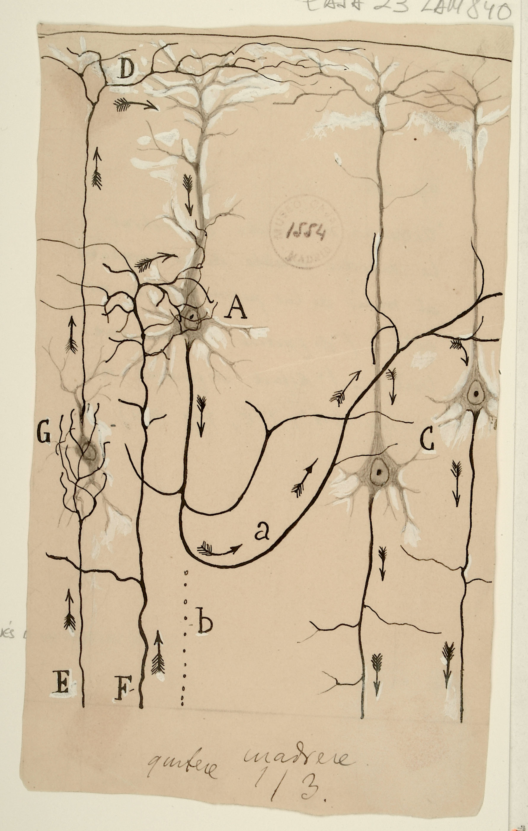
Scheme to explain the mechanism of vomiting and coughing (1904)

Div
classgrid-row grid-gap
Div
classdesktop:grid-col-6
Div
classdesktop:grid-col-6
Span
classcredit

Courtesy of the Cajal Institute, Spanish National Research Council or CSIC©

Div
classgrid-row grid-gap
Div
classdesktop:grid-col-6

This diagram details the body-spanning circuit underlying cough and vomiting.  Irritation of sensory neurons in the larynx (A) or the stomach (J) initiates signals that travel through the vagus nerve (B) to pre-motor neurons (M, D), and onwards to motor neurons (F,H) that then control the movement of the chest and abdominal wall muscles (K) or the smooth muscle of the stomach (I), respectively.  The cough reflex is extremely important for clearing the lungs of impediments that inhibit their gas-exchanging function. Its dysregulation can lead to damage of the airway mucosa and degrade quality of life.  New targets for cough-suppressant drugs are being sought in the sensory neurons in the lungs and throat (A) and include blockers of transmembrane channels aimed at reducing nerve excitability.

Div
classdesktop:grid-col-6

Image Added

Span
classcredit

Courtesy of the Cajal Institute, Spanish National Research Council or CSIC©


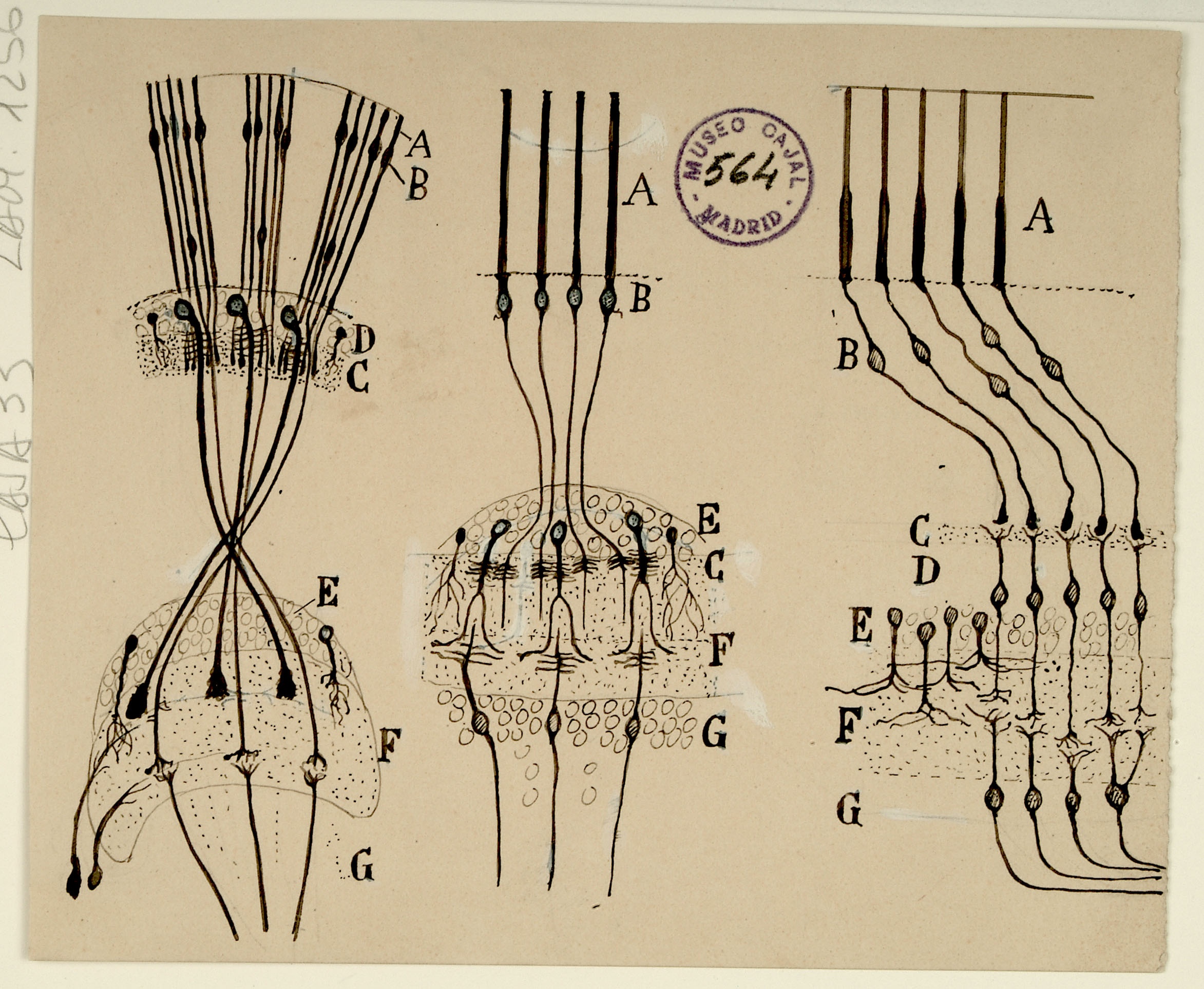
Schemes intended to facilitate the comparison between retinas of insects, cephalopods and vertebrates

Div
classgrid-row grid-gap
Div
classdesktop:grid-col-6

Cajal’s fascination with the retina is well documented – his publications on retinal neurons and organization in various species span 45 years, the length of his career.  Shown above are schematics of insect (left), cephalopod (middle), and vertebrate (right) retinas, highlighting their similarities.  All of these retinas are composed of photoreceptors (A) which are activated by light and transmit signals through synapses of intermediary cell types (B, C, F) onwards toward deeper brain processing (G).

Cajal has oriented all these diagrams with the photoreceptors at the top for ease of comparison, but in fact the diagram of the vertebrate retina is more correctly shown inverted compared to the other two, with its photoreceptors located at the bottom of the diagram.  This is because the photoreceptors of invertebrates, such as insects and cephalopods, are exposed to light directly as the top layer of the retina, while the photoreceptors of vertebrates are actually at the bottom of the retina. Light must pass through the transparent layers of nerves above them before reaching them.

Div
classdesktop:grid-col-6

Image Added

Span
classcredit

Courtesy of the Cajal Institute, Spanish National Research Council or CSIC©


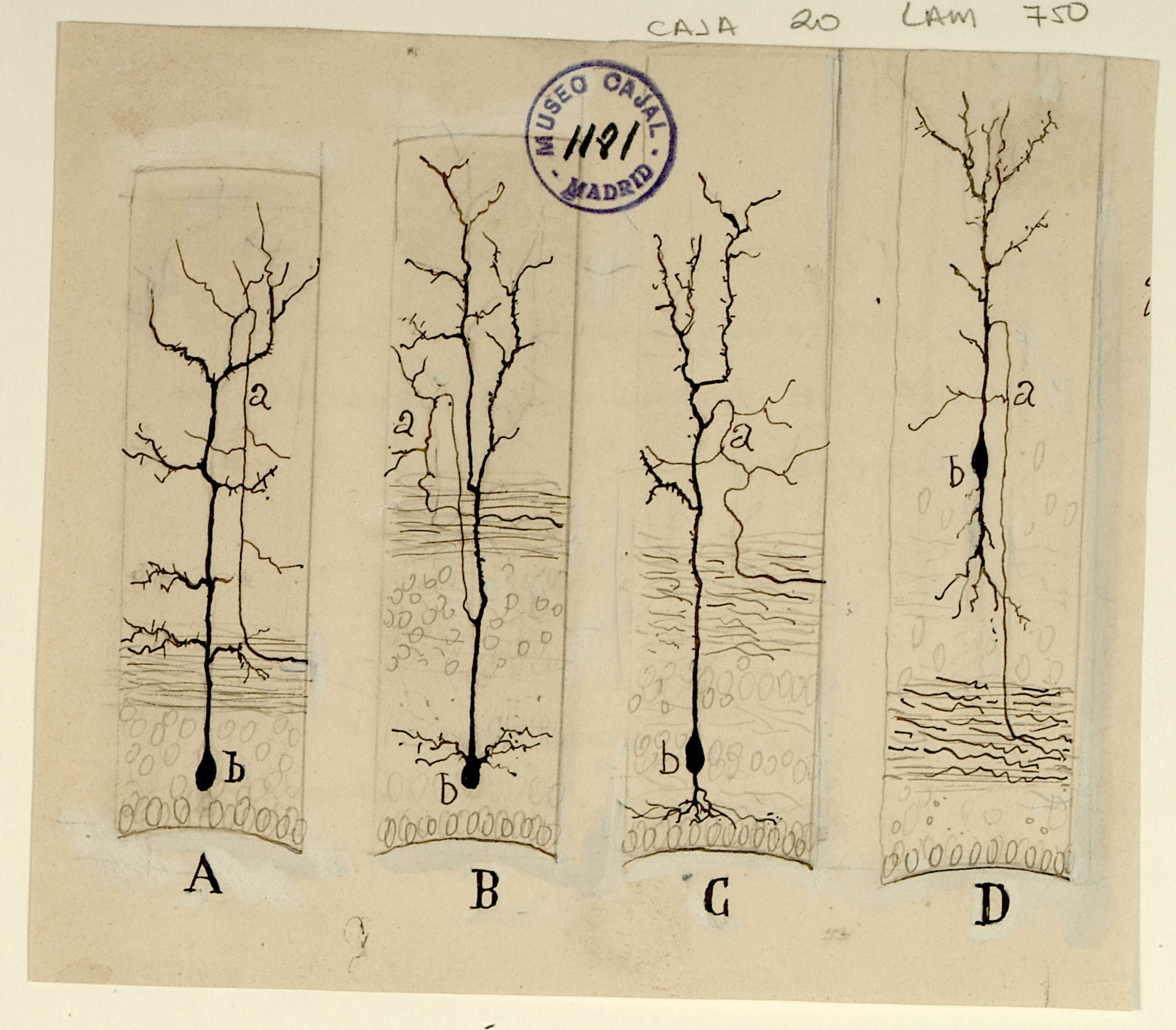
Possible course of information flow through the pyramidal neurons in the cerebral cortex

Div
classgrid-row grid-gap
Div
classdesktop:grid-col-6

Here, Cajal illustrates a possible flow of information within the cerebral cortex, focusing on 4 pyramidal neurons.  Inputs (E,F) come in several forms, including projections to apical dendrites (D) and to intermediate layers (A). After relay through other cortical neurons (a), deeper layer neurons (C) project out of the cortex. While there is strong anatomical evidence for these canonical paths of information flow, in recent years we have learned that the high degree of connectivity and diversity of cells in the cortex makes the fundamental computation of the cortex quite complex: a single cortical column has hundreds of thousands of such pyramidal cells, connected by millions of synapses. Cajal’s depiction of this circuit presages the 'canonical cortical microcircuit' of Douglas and Martin by nearly 100 years.  The field is currently engaged in an effort to extend the canonical circuit ideas to the entire cortical network: understanding how the massive local connectivity builds on this canonical circuit backbone to create cortical computation.

Div
classdesktop:grid-col-6
Span
classcredit

Courtesy of the Cajal Institute, Spanish National Research Council or CSIC©

Divclassgrid-row grid-gap

Div
classdesktop:grid-col-6
Div
classdesktop:grid-col-6

Image Added

Span
classcredit

Courtesy of the Cajal Institute, Spanish National Research Council or CSIC©

6th Installation

...

Motor cortex

Div
classgrid-row grid-gap
Div
classdesktop:grid-col-6

Formerly known as the ascending frontal gyrus, the precentral gyrus is the home of the primary motor cortex, which works in association with other motor areas to plan and execute movement.  The primary motor cortex contains a variety of pyramidal neurons whose axons extend down the spinal cord and synapse with motor neurons.  Shown above are pericellular nests around pyramidal cell bodies (b,c,d) in the primary motor cortex, formed by the profusely branched axons of other neurons. Cajal noted the presence of pericellular nests in the cerebellum, hippocampus, and cortex and hypothesized that they amplify signals from the presynaptic cells to the enveloped cell body.  We now know that pericellular nests allow tight control of the targeted cell, crowding out input from other, more distant axons.  While the precise function of the pericellular nests above remains unknown, recent research in the hippocampus has shown that they can influence memory and reinforcement of learned behaviors.

Button

Div
classdesktop:grid-col-6

Illustration

Span
classcredit

Courtesy of the Cajal Institute, Spanish National Research Council or CSIC©

...