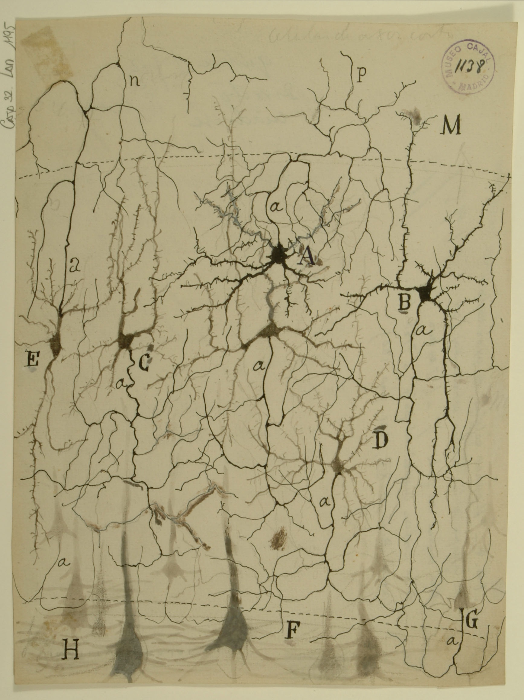
The primary visual cortex is the first processing center for visual signals within the brain, which is divided into six functionally distinct layers. Shown here are feline stellate cells (A,B) from Layer 4 of the primary visual cortex. Stellate cells receive the majority of their input from the lateral geniculate nucleus (LGN) of the thalamus, then send signals to pyramidal cells in layer 6 (H,F) for further processing. While studies of visual system gross anatomy dating from the 1940s indicated that information travels from the eye through the LGN and then into the cortex, more recent work has revealed that only 5-10% of the excitatory synapses onto stellate cells actually come from the LGN; most input comes from deeper cortical layers. The mechanism by which so few LGN inputs control stellate cells remains unknown – synapses from the LGN not appreciably larger than the others, nor are they clustered on stellate cell dendrites. One possible explanation is that signals from the LGN are highly synchronized with each other, whereas other inputs are more temporally dispersed.
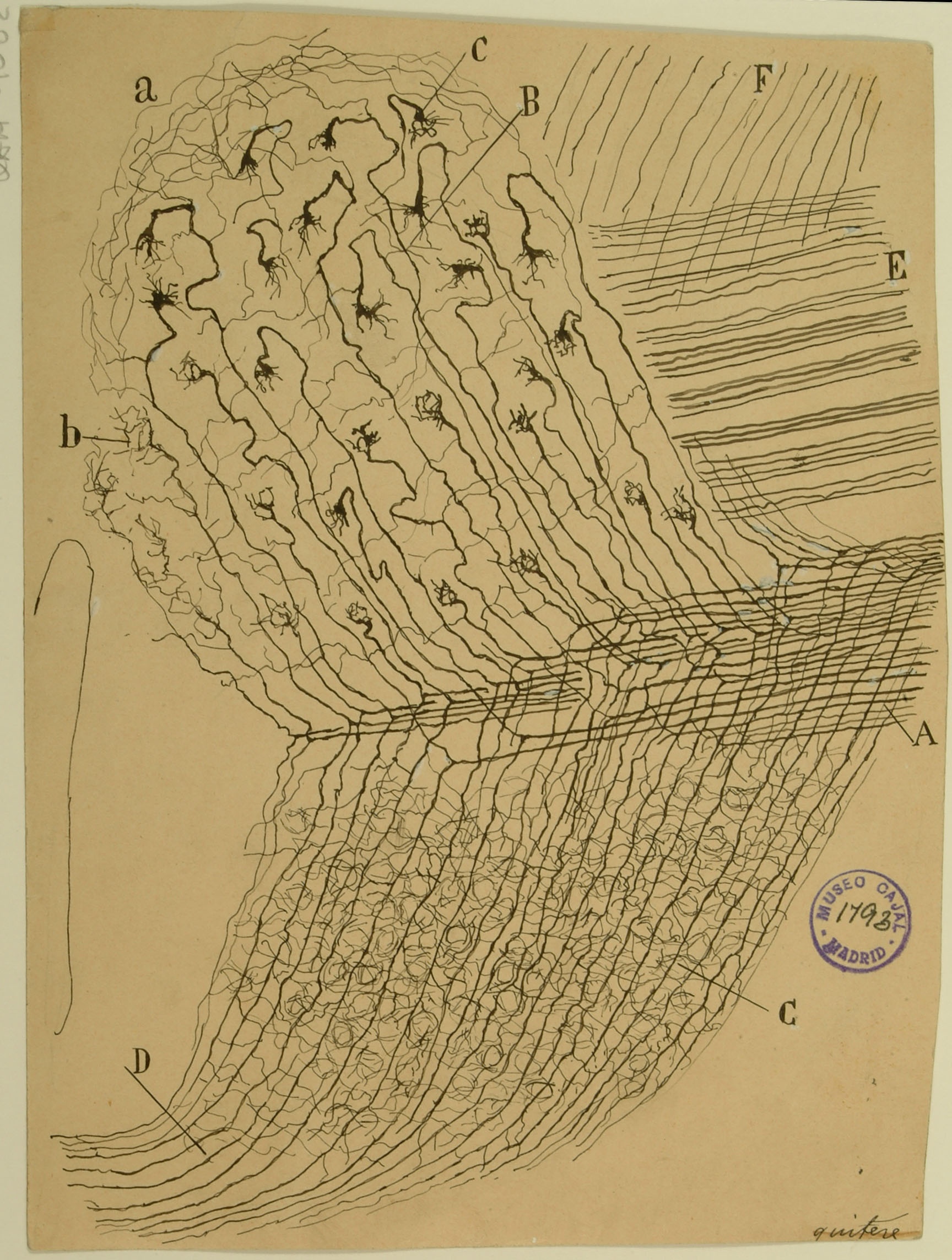
The ventral cochlear nucleus is the first processing center for auditory signals in the central nervous system. Here, Cajal shows how individual auditory nerve fibers enter the cochlear nucleus ventrally as the cochlear nerve (A), bifurcating to form ascending and descending branches. The ascending branches (B) give rise to complex axonal terminals, the Calyces of Held, which synapse onto bushy cells to facilitate rapid, high-fidelity synaptic signals crucial to precise transmission of acoustic information. The descending branches travel posteriorly, giving rise to synapses in both the ventral (C) and dorsal (D) cochlear nucleus subdivisions that will perform more complex auditory processing, integrating feedback signals from deeper parts of the auditory pathway together with input from the cochlear nerve.
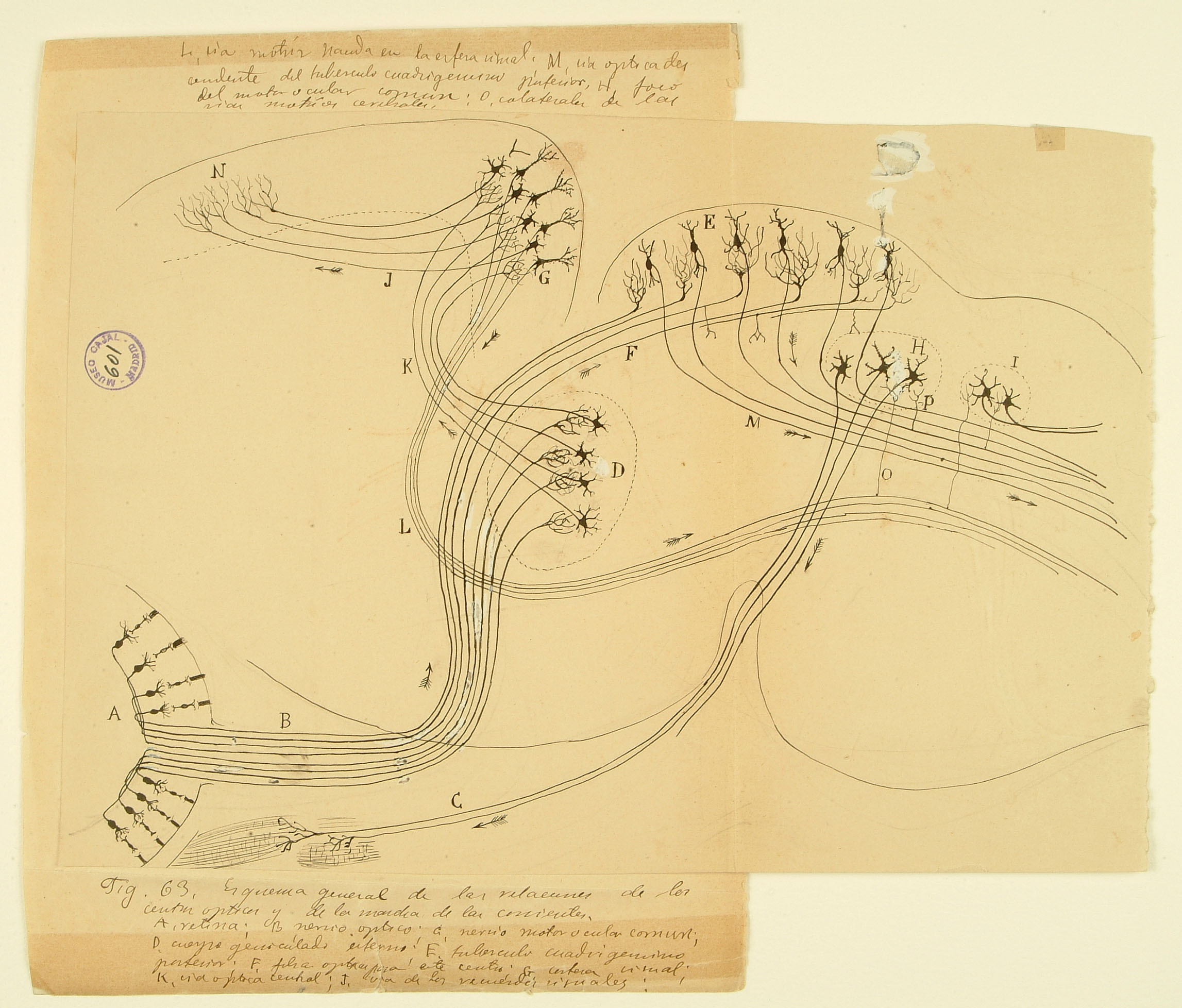
This drawing of the rodent early visual system illustrates one of Cajal’s most important contributions to science: the law of dynamic polarization, wherein information flows from the receptive dendrites of a neuron, through its cell body, and down through its axon, which then makes synaptic contacts onto dendrites of subsequent neurons in the pathway. Here he shows two routes by which visual information is used to control movements of the body. Signals from the retina (A) enter the brain by the optic nerve (B) and terminate in two different brain regions – the lateral geniculate nucleus of the thalamus in the forebrain (D) and the superior colliculus on the roof of the midbrain (E). The superior colliculus, evolutionarily conserved across mammal, integrates visual and other inputs to provide signals (M) instructing physical movement and orientientation. Neurons in the visual thalamus (D) relay signals to the visual cortex (G), which sends projections to higher-order cortical areas (N) and also provides another set of descending motor commands (L). This diagram was submitted by Cajal as part of a joint application for the 1902 Martinez y Molina Award titled “Sensory Centers in Man and Animals” with his brother Pedro, also a noted histologist who specialized in amphibian and reptilian brain structures.