
...
Peter Sonderegger, while a post-doc at the National Institute of Child Health and Development (NICHD), used the GELLAB-I system with the RTPP to investigate how the expression of axonal proteins of sensory and motor neurons was influenced by non-neuronal cells [27, 29, 45]. At the same time we were investigating the feasibility of porting GELLAB-I (written in SAIL) to the PASCAL computer language. The inflexibility of PASCAL eventually led us to convert GELLAB-I to the portable C/UNIX/X-windows environment called GELLAB-II [42, 43, 44, 45] The DECsystem-10 SAIL version, GELLAB-I, was exported to research labs at Univ. of Chicago (Eric Lester) and Univ. of Kiel (Heinz Busse). The Unix version, GELLAB-II, was exported to a number of research labs around the world (CDC with Jim Myrick [44], Univ. Zurich with Peter Sonderegger, Agr. Univ. Norway with Trygve Krekling, and others) and led to a commercial subset version for Windows PCs called GELLAB-II++ by CSPI/Scanalytics.
| Anchor | ||||
|---|---|---|---|---|
|
...
Partly as a result of this collaboration, Jake became more interested in computing, which led him to purchase one of the early DEC VAX computers for his lab. As Jake became more involved with computational techniques, he realized the general importance, as did Lew, of using computers in biology. Eventually, this interest culminated with the National Cancer Institute's purchase of a Cray XMP supercomputer, and the establishment of what is today the Advanced Biomedical Computer Center in Frederick. The Cray XMP, at that time, was the first supercomputer in the world solely dedicated to biomedical research. Thus, the impact of the RTPP continues to be felt in the biological computation sciences 30 years later.
| Anchor | ||||
|---|---|---|---|---|
|
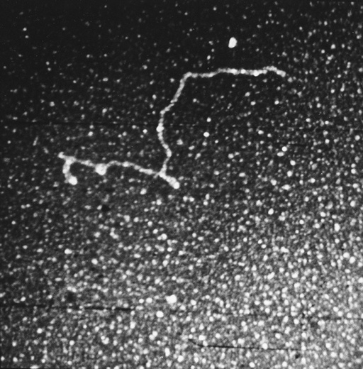
Figure 20. One of the early RNA electron micrographs scanned with the vidicon/RTPP system (Jacob Maizel, Bruce Shapiro, and Lewis Lipkin) [33, 34, 39, 48, 49]. The sample was adenovirus type 2 messenger RNA. Bruce developed boundary segmenters and boundary shape descriptors that could map electron micrograph data to the secondary structure.
...
Overview
Content Tools
ThemeBuilder