On March 31, 2014, the second phase of the John Edward Porter Neuroscience Research Center was dedicated. This new facility is shared by scientists from the National Institute of Neurological Disorders and Stroke (NINDS), National Eye Institute (NEI), National institute of Child Health and Human Development (NICHD), National Institute of Dental and Craniofacial Research (NIDCR), National institute of Mental Health (NIMH), National Institute on Deafness and Other Communicable Disorders (NIDCD), and the National Institute of Biomedical Imaging and Bioengineering (NBIBNIBIB)—and represents a unique opportunity for scientists to collaborate across academic disciplines as well as the boundaries that sometimes separate institutes and centers on the NIH campus.
A new wave of research and exploration is beginning within these walls with new support for the creation of a new arsenal of instruments for unlocking the mysteries of the brain through the Brain Research through Advancing Innovative Neurotechnologies (BRAIN) Initiative. This moment may be the most appropriate to look back over the accomplishments of the last century and anticipate those of the next.
Table of Contents
maxLevel
2
minLevel
2
Div
class
desktop:grid-col-6
Center
Span
class
caption
This Exhibit currently located in building 35
...
Div
class
grid-row grid-gap
Div
class
desktop:grid-col-6
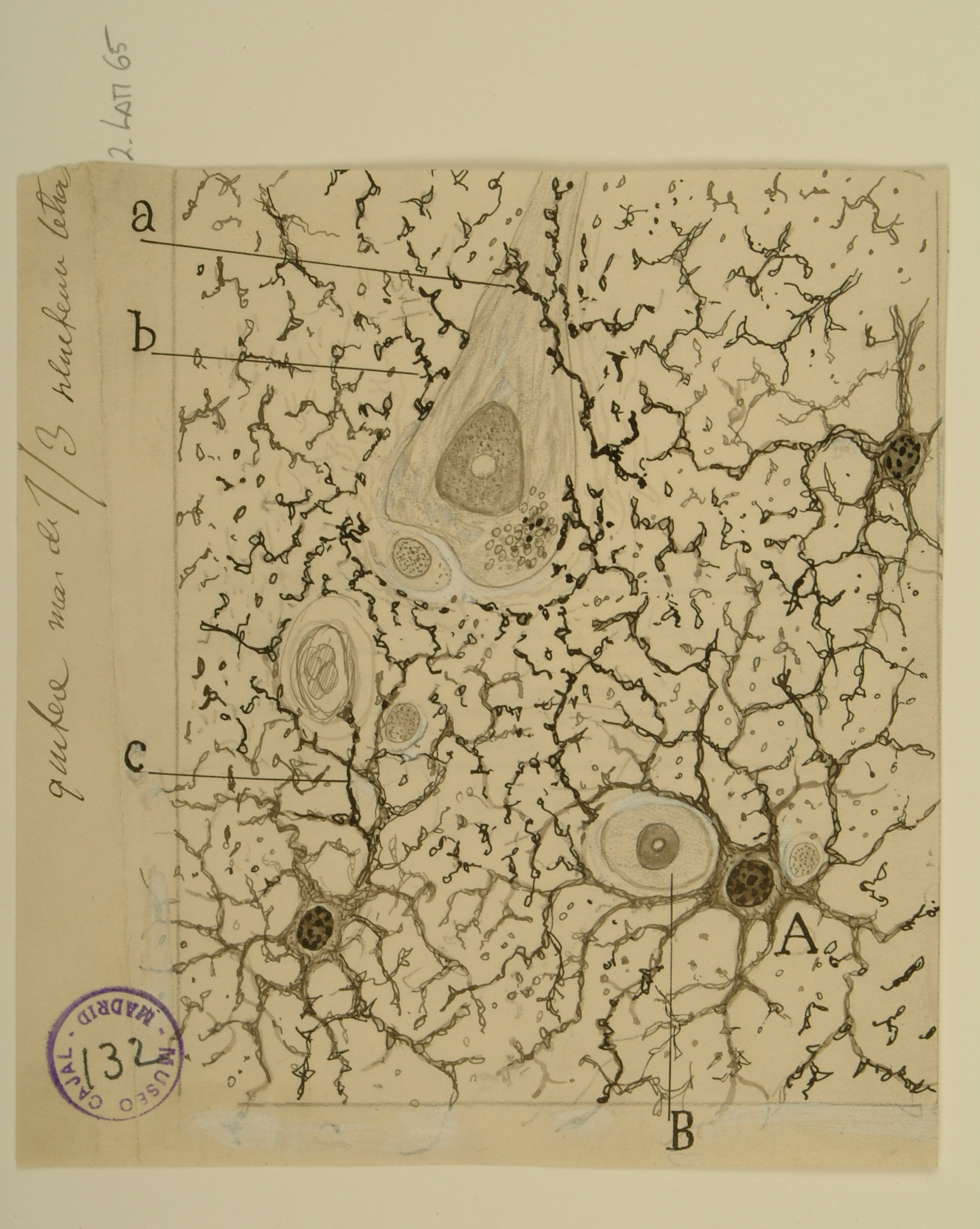
Once thought to be mere “filler” for the space around neurons, astrocytes are star-shaped glial cells found throughout the brain and spinal cord that are now known to perform many important functions, such as regulating the transmission of ions and glucose between blood vessels and the brain.
When Cajal drew these protoplasmic astrocytes, the prevailing theory was that astrocytes only provided structural support for neurons. He rejected this idea and instead hypothesized that all the astroglia in the brain formed a sort of gland, which would release substances that affect brain function; it is now known that astroglia do indeed release substances that affect neuronal signaling, including glutamate, GABA, and ATP.
Having observed the close association of astrocytes with blood vessels and neurons, as well as the fact that all astrocytes appeared to have a prominent appendage, or “foot,” Cajal further hypothesized that astrocytes used this “foot” to stimulate blood vessel dilation. Astrocyte “feet” do indeed regulate blood vessel diameter, albeit via the release of signaling molecules rather than through physical manipulation, as Cajal imagined. Astrocyte-evoked changes in blood flow, and thus in oxygenation, constitute the signal that is measured by fMRI, a tool used to image brain activity.