
The Ultimate Portrait Painter
Howard Bartner and 40 Years of Medical Science
Introduction
Since the sixteenth century, when Andreas Vesalius' (1514-1564) drawings of dissections revolutionized our knowledge of human physiology, medical illustrators have become the ultimate portrait painters, rendering the essence of human life. To this day, medical illustrations remain the primary source of information for students of human biology and medicine. Continuing in the tradition of Vesalius, Howard Bartner of the Medical Arts and Photography Branch at the National Institutes of Health has devoted forty years to portraying human anatomy in his drawings. This exhibit looks at only a fraction of his work.
 Illustrator Howard Bartner
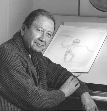
Born in New York City in 1931, Howard Bartner discovered his artistic abilities early, painting landscapes and still lifes for his aunts and uncles. After graduating from the Stella Elkins Tyler School of Fine Arts at Temple University, Bartner found that medical illustration melded his interest in biology and the sciences with his artistic talent.
As a student in the medical arts graduate program at the Johns Hopkins University School of Medicine, Bartner's first encounter with the cadaver was difficult, but the intricate beauty of the human body fascinated him. Now he draws his illustrations from original dissections, stopping periodically to sketch, or observing surgical procedures and examining patients. Bartner's direct observations often produce works of considerable artistic sensitivity and beauty.
In addition to his career at the National Institutes of Health, Bartner is also an Assistant Professor in the Department of Art as Applied to Medicine at the Johns Hopkins University School of Medicine.
Ophthalmological Disorders
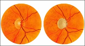
In this series of paintings of the inside of the eye, the orange circle represents the retina and the yellow circle represents the optic disc, which is where the optic nerve that connects the eye to the brain leaves the retina.
Painting 1: Glaucomatous Optic Disc
In the normal optic disc on the left, the number of optic nerve fibers is normal. The cup within the optic nerve is small and the vessels are near the center of the disc. In the glaucomatous optic disc on the right, increased pressure within the eye has caused the disappearance of a large number of optic nerve fibers. Therefore, the cup has enlarged and the disc vessels have curved along the cup's contour.
Painting 01
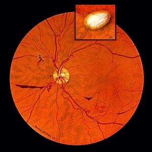
Painting 2: Retina Uveitis Hemorrhages
In 1958, Howard Bartner created his first painting at the National Institutes of Health: this view of the retina of a patient with uveitis, an inflammation of the uvea (iris, ciliary body, and choroid), the middle layer of the eye. As a result, the retinal vessel walls are weakened and are bleeding. The inset depicts a scar in the retina; the retina and choroid have disappeared and the whiteness of the sclera (the outer layer of the eye) can be seen.
Painting 02
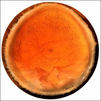
Painting 3: Detached Retina
This painting, completed in 1967, shows an entire retina. The optic disc is the small yellow circle near the center of the orange retina. The billowing of the bottom segment of the retina indicates that this segment of the retina has become detached from the firm connective tissue that encloses the eye. This causes visual loss in the area.
Painting 03
Anatomy of the Infant Head
Because infant anatomy of the head differs greatly from adult cranial anatomy, Dr. James Bosma of the National Institute of Dental and Craniofacial Research wanted to produce a reference guide on infant anatomy for pediatricians, pediatric dentists, maxillofacial surgeons, and other physicians. Bosma hoped that by learning more about the development of the infant airway, throat, larynx, and pharynx, that doctors could better understand breast feeding and respiratory abnormalities. Bosma asked Howard Bartner to collaborate with him and three other illustrators in the study and description of the infant head, resulting in the publishing of Anatomy of the Infant Head (Johns Hopkins University Press, 1986). Bartner's elegant illustrations are important features of this classic text.
Below are side-view drawings of an infant's head. Note the nugget of extra fat, known as the "buccal pad." The buccal pad contributes to babies' chubby cheeks and diminishes when the infant is about a year old. Drawings two and three depict arrangements of the jaw and throat muscles.
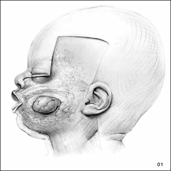
Drawings 1-3: Sequential Dissections of the Head from the Side
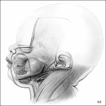
Drawing 2 of 3: Sequential Dissections of the Head from the Side
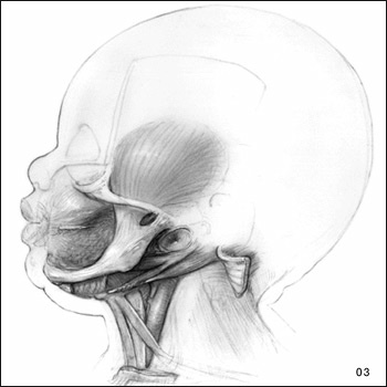
Drawing 3 of 3: Sequential Dissections of the Head from the Side
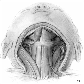
Sequential Views of the Infant's Throat, Larynx Areas, and Mouth Muscles from the Front
Removal of the skin shows tissue and muscles in the throat area. Note how the drawings become progressively more detailed as the levels of dissection go deeper. The left side of drawing 6 is a working sketch; the finished product is on the right.
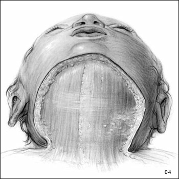
Drawing 1 of 4: Sequential Views of the Infant's Throat, Larynx Areas, and Mouth Muscles from the Front
Drawing 2 of 4: Sequential Views of the Infant's Throat, Larynx Areas, and Mouth Muscles from the Front
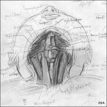
Drawing 3 of 4: Sequential Views of the Infant's Throat, Larynx Areas, and Mouth Muscles from the Front working sketch.
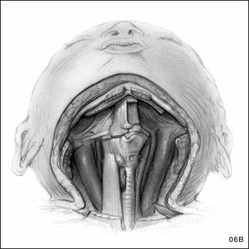
Drawing 4 of 4: Sequential Views of the Infant's Throat, Larynx Areas, and Mouth Muscles from the Front.
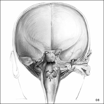
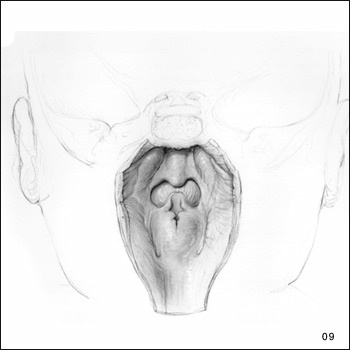
Drawings 7-9: Sequential Drawings of the Throat Area from the Back
The following drawings show the back of an infant's head. Drawing seven depicts the relation between the pharynx and vessels and nerves on each side of it. The eighth drawing illustrates the ear as seen from the back; and the ninth drawing is a close-up of the pharynx and larynx areas.

Drawing 1 of 3: Sequential Drawings of the Throat Area from the Back
Drawing 2 of 3: Sequential Drawings of the Throat Area from the Back
Drawing 3 of 3: Sequential Drawings of the Throat Area from the Back
Human Physiology in Space
In some of his most recent work, Bartner created illustrations for the textbook Human Physiology in Space, produced jointly by the National Aeronautics and Space Administration (NASA), NIH, the Universities Space Research Association, and the University of Texas Southwestern Medical Center. The textbook uses space science to inspire high school students to learn about human physiology. Bartner invented two characters, a teenage boy and girl, to demonstrate the ideas in the text; his whimsical drawings keep students involved.
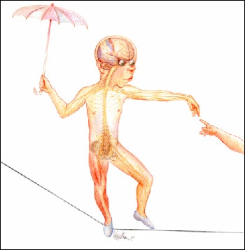
Drawing 1: Boy on a Tightrope
Bartner's drawing of a teenage boy walking a tightrope shows how the body's sensory organs work together with the bones and muscles to maintain balance, keeping the boy from falling off the rope. Bartner incorporated Michelangelo's Sistine Chapel painting of Adam's birth at God's hand (right side of the drawing). "For me, the drawing also suggests the fragility of life," says Bartner.
Drawing 2: The Inner Ear
In this series of drawings, Bartner shows in increasing detail the vestibule organs of the inner ear responsible for balance. The three curved canals of the inner ear detect when the head rotates, and the two small organs they join (the otolith organs) detect when the head moves linearly. That's why an infection in your inner ear may affect your balance.
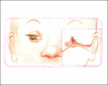
Drawing 1 of 3: Details of the vestibule organs of the inner ear responsible for balance
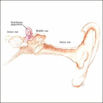
Drawing 2 of 3: Details of the vestibule organs of the inner ear responsible for balance
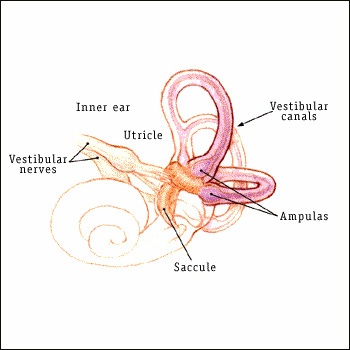
Drawing 3 of 3: Details of the vestibule organs of the inner ear responsible for balance
Photography Credits
All images, text, transcription, and/or recordings reproduced in these exhibits and galleries may be protected by copyright. Users who wish to reproduce materials for other than private use should make an additional effort to determine ownership and, if restricted, seek permission before reproducing them.
Related Links
- Johns Hopkins University Medical Arts Program
- National Eye Institute
- Glaucoma Research Foundation
- Johns Hopkins University Press
- National Institute of Child Health and Human Development
- National Institute of Dental and Craniofacial Research
- National Aeronautics and Space Administration
- Universities Space Research Association
- University of Texas Southwestern Medical Center
Overview
Content Tools
ThemeBuilder